. 2025 Aug 12;16(1):7435.
doi: 10.1038/s41467-025-62788-6.

A citrullinated histone H3 monoclonal antibody for immune modulation in sepsis

Affiliations

A citrullinated histone H3 monoclonal antibody for immune modulation in sepsis

Wenlu Ouyang et al. Nat Commun. .

Abstract

Citrullinated histone H3 (CitH3), released from immune cells during early sepsis, drives a vicious cycle of inflammation through excessive NETosis and pyroptosis, causing immune dysfunction and tissue damage. To regulate this process, we develop a humanized CitH3 monoclonal antibody (hCitH3-mAb) with high affinity and specificity to target this process. In murine models, hCitH3-mAb reduces cytokine production, mortality and acute lung injury (ALI) caused by LPS and Pseudomonas aeruginosa while enhancing bacteria phagocytosis in the lungs, spleen, and liver. Using pre-equilibrium digital ELISA (PEdELISA), we identify an optimal therapeutic window for hCitH3-mAb in sepsis-induced ALI. In parallel, we explore the molecular mechanism underlying CitH3-driven inflammation. We find that in macrophages, CitH3 activates Toll-like receptor 2 (TLR2), triggering Ca2+-dependent PAD2 auto-citrullination and nuclear translocation, amplifying CitH3 production via a harmful feedback loop. The hCitH3-mAb treatment effectively disrupts this cycle and restores macrophage function under septic conditions. Together, these findings highlight both the therapeutic potential of hCitH3-mAb and provide a deep mechanistic insight into the CitH3-PAD2 axis in sepsis, supporting its further development for treating immune-mediated diseases.


Conflict of interest statement

Competing interests: Patent application related to the hCitH3-mAb technology was filed by University of Virginia Patent Foundation, The Reagents of University of Michigan and HTIC, Inc. J.M., Y.L. and T.T. are co-founders of HTIC that develops hCitH3-mAb for immune modulation applications. The remaining authors declare no competing interests.

Figures

Fig. 1
Fig. 1. Thematic approach for the optimization and scale-up production of hCitH3-mAb.
a Schematic flowchart illustrating the humanization process of mouse CitH3-mAb and the optimization steps conducted during developability studies, culminating in the scale-up production of hCitH3-mAb. b Plasmid map detailing the co-expression of the heavy chain and light chain of hCitH3-mAb in CHO cells. c Quantification of cell density for different stable CHO clones expressing hCitH3-mAb. d Assessment of cell viability across the various CHO clones stably expressing hCitH3-mAb.e, Titer quantification of stable CHO clones, highlighting clone P2D6, which exhibited the highest hCitH3-mAb titers on days 13 and 14. Clone P2D6 was selected for harvesting and purification. f RF-HPLC analysis demonstrated a homogeneous distribution of the purified hCitH3-mAb, achieving >99.5% purity (dominant peak) with no detectable aggregation. g SDS-PAGE analysis of purified hCitH3-mAb revealed a distinct heavy chain and light chain under reducing conditions, and a single band under non-reducing conditions, indicating high purity and proper assembly. For every batch of the hCitH3-mAb, such quality control assessment is routinely performed by CDMO. Similar results were obtained from ≥3 independent replicates.Source data are provided as a Source Data file.
Fig. 2
Fig. 2. hCitH3-mAb exhibits superior binding capacity to CitH3 compared to commercial CitH3 antibodies.
a Indirect ELISA quantification demonstrating the sensitivity of hCitH3-mAb (blue) versus commercial CitH3-mAb-3Cit antibodies (black). CitH3 standards and test samples were identically diluted, and ELISA procedures were performed in parallel under identical conditions (n = 3). Statistical significance was analyzed using two-way ANOVA (two-sided). ***p < 0.001, ****p < 0.0001. The asterisks in the figure indicate comparisons between hCitH3-mAb and commercial CitH3-mAb-3Cit antibodies. b Western blot analysis comparing the sensitivity and specificity of hCitH3-mAb and CitH3-mAb-3Cit. Four peptides (H3, CitH3 (R2,8,17,26), AceH3, and MetH3)were each loaded at 0.5 µg. After electrophoresis and membrane transfer, the blot was divided and probed separately with either hCitH3-mAb or CitH3-mAb-3Cit (2 µg/L). Membranes were processed and exposed simultaneously (n = 3 independent experiments). Data are presented as mean ± SD. Statistical analysis was performed using a two-sided t-test (**p < 0.01). c ELISA of human septic serum samples, comparing the sensitivity of hCitH3-mAb and CitH3-mAb-3Cit. d ELISA quantification of CitH3 protein levels in human serum samples. Serum was collected from patients at enrollment, at 24- and 48 h post-enrollment. For the infectious groups, n = 6 at both 24- and 48 h; for the other group, n = 5. The ‘Mild’ group corresponds to patients with a total SOFA score ≤6, while the ‘Moderate-to-Severe’ group corresponds to a SOFA score >6. Non-infectious controls represent patients who experience shock without clinical or laboratory evidence of infection. Data are presented as mean ± SD. Statistical significance was determined using two-way ANOVA with Turkey’s multiple comparisons test (*p < 0.05, **p < 0.01).Source data are provided as a Source Data file.
Fig. 3
Fig. 3. hCitH3-mAb reduces inflammation and protects against sepsis-induced ALI in mice.
a Kaplan-Meier curves of mice after administration of Pseudomonas aeruginosa (P. aeruginosa, 2.5 × 10⁶ CFU) for 10 days. Mice were divided into two groups: one receiving hCitH3-mAb (20 mg/kg) and the other receiving human IgG, with treatments administered within 30 min of sepsis onset. Data represent pooled results from three independent experiments (total n = 22; 6–8 mice per group per experiment). b Bacterial load in the lung, spleen, and liver 24 h after P. aeruginosa inoculation in mice treated with either hCitH3-mAb or human IgG (n = 3 per group). Homogenized organs were weighed, serially diluted, and plated on nutrient agar plates. Colony counts were obtained using ImageJ after 16-hour incubation at 37 °C. Data are presented as mean ± SD. c Levels of CitH3, IL-1β, and IL-6 in bronchoalveolar lavage fluid (BALF) from mice treated with hCitH3-mAb, compared to those treated with PBS or human IgG (n = 4 per group). Data are presented as mean ± SD. d Representative images and assessment of lung tissue sections stained with H&E. ALI scores and the proportion of airspace area were quantified by a blinded pathologist (n = 4 per group). Data are presented as mean ± SD. Statistical analyses were performed using Kaplan-Meier analysis with two-sided log-rank test for (a), two-sided t-tests for (b, d), and one-way ANOVA with Dunnett’s multiple comparisons test for (c): *p < 0.05, **p < 0.01, ***p < 0.001, ****p < 0.0001.Source data are provided as a Source Data file.
Fig. 4
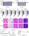
Fig. 4. Safety profile of CitH3-mAb in C57BL6 mice and negative ADCC effect of hCitH3-mAb.
a Body weight of mice before and during the treatment for 6 weeks. Mice were divided into two groups, 9 receiving CitH3-mAb (10 mg/kg) and the other 9 receiving saline, with treatment administrated twice per week (n = 9 biological samples per group). b Ejection fraction (EF) data of mice treated with CitH3-mAb (10 mg/kg) or saline. Echocardiography assay were conducted before and every two weeks during the treatment (n = 9 biological samples per group).c, Levels of BUN, total protein, globulin, albumin/globulin ratio, ALT, ALP and non-fasting glucose in serum from mice treated with CitH3-mAb, compared to those treated with saline. Creatinine of all the indicated mice were below 0.4 mg/dL. 4 out of 9 mice in each group were randomly selected for serum chemistry assay and histological analysis (n = 4 biological samples per group)d, Representative images and assessment of vital organ sections (heart, lung, liver, kidney, spleen) stained with H&E (n = 4 per group). e ADCC evaluation of hCitH3-mAb in THP-1 cells. PD-L1 transient expression in THP1 cells was used as positive control for ADCC. No ADCC response was observed with hCitH3-mAb (n = 2 per group; GLP-compliant study at a certified CRO). f Pharmacokinetics of hCitH3-mAb in Cynomolgus monkeys following intravenous infusion. Two doses of hCitH3-mAb (30 or 200 mg/kg) were administered, and the plasma concentrations of the antibody were quantified during the one-week observation time. The estimated half-life was 76.7 and 47.7 h, for 30 mg/kg and 200 mg/kg dosing, respectively. (n = 2 per dose group; GLP-compliant study at a certified CRO) Data are presented as mean ± SD. Statistical analysis was performed using two-sided t-tests: ns no significance. Source data are provided as a Source Data file.
Fig. 5
Fig. 5. hCitH3-mAb protects against LPS-induced endotoxemia in mice by mitigating cytokine storm development.
a Kaplan-Meier curves of mice monitored over 10 days following lipopolysaccharide (LPS, 25 mg/kg) administration. Septic mice received hCitH3-mAb (20 mg/kg) or human IgG (20 mg/kg) via tail vein injection 30 min after sepsis onset. Data represent pooled results from three independent experiments (total n = 24; 8 mice per group per experiment). b Illustration of traditional ELISA and PEdELISA for the detection of cytokines and CitH3. c Blood samples were collected at 3 h intervals from the same mouse (n = 3 per group) and analyzed using PEdELISA. 8 µL of serum samples were used for CitH3 quantification, revealing a biphasic response of LPS-induced CitH3 elevation indicative of sepsis onset and cytokine storm development. hCitH3-mAb treatment mitigated LPS-induced CitH3 elevations. Data are presented as mean ± SD. d Serum levels of IL-1β, IL-6, and TNFα in the LPS-induced endotoxemia model (n = 3 per group). Statistical analysis was performed using multiple t-tests: *p < 0.05, **p < 0.01, ***p < 0.001. Data are presented as mean ± SD.Statistical analysis was performed using Kaplan-Meier analysis with two-sided log-rank test for (a), two-sided multiple t-tests for (c, d). *p < 0.05, **p < 0.01, ***p < 0.001, ****p < 0.0001.Source data are provided as a Source Data file.
Fig. 6
Fig. 6. Bacteria-induced injury to macrophages is mitigated by hCitH3-mAb.
a Immunoblot analysis of CitH3 levels in the supernatant (s.n.) and cell pellets of THP-1 cells pre-treated with hCitH3-mAb or human IgG (1.5 μg/mL), followed by exposure to P. aeruginosa (MOI 100) for 2 or 4 h. Similar results from three independent replicates. b Viability of THP-1 cells treated with P. aeruginosa at MOI 100 for 2 h with increasing doses of hCitH3-mAb or IgG (n = 5). c Phagocytic ability of THP-1 macrophage cells treated with hCitH3-mAb or human IgG (1.5 μg/mL) for 2 h, followed by P. aeruginosa exposure (MOI 100, 1 h) and incubation with pHrodo Red E. coli BioParticles (0.1 mg/mL). In representative images, red fluorescence indicates phagocytic activity, and blue signals represent Hoechst-stained nuclei. Fluorescence was measured at 560/585 nm (n = 6 biological replicates). Statistical analysis was performed using one-way ANOVA with Tukey’s multiple comparisons test. d Cell death in THP-1 macrophages incubated with BALF for 4 h. BALF was collected from P. aeruginosa-induced septic mice pre-treated with either hCitH3-mAb (20 mg/kg) or human IgG (20 mg/kg) (n = 6 per group). BALF was collected in RPMI 1640 medium and filtered through a 0.22 μm filter. BALF was added at 12.5% of the final culture volume. e Levels of IL-6, IL-1α, IL-1β, and TNFα in the supernatant of THP-1 macrophages (n = 6 biological replicates) infected with P. aeruginosa (MOI 100, 2 h). NC indicates the negative control group. BALF was added at 12.5% of the final culture volume. f Western blot analysis of pyroptosis markers, including Caspase-1 and GSDMD cleavage products, in BMDMs treated with BALF as described in (e). hCitH3-mAb treatment was more effective than the commercial CitH3-mAb-3Cit in mitigating BALF-induced pyroptosis in BMDMs (n = 4 biological replicates). Human IgG was used as negative control. g Levels of IL-1α, IL-1β, IL-18, and LDH in the supernatant of BMDM cells treated with BALF. hCitH3-mAb demonstrated greater efficacy than commercial CitH3-mAb-3Cit in reducing BALF-induced pro-inflammatory cytokine release in BMDMs (n = 6 biological replicates).Data are presented as mean ± SD. Statistical analyses were conducted using one-way ANOVA and Dunnett’s test, with comparisons among the BALF-treated group for (d–g), and to the ‘0’ group for (a, b): *p < 0.05, **p < 0.01, ***p < 0.001, ****p < 0.0001.Source data are provided as a Source Data file.
Fig. 7
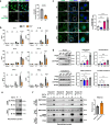
Fig. 7. Circulating CitH3 triggers Ca²⁺-dependent PAD2 auto-citrullination and nuclear translocation via TLR2 signaling.
a Immunofluorescence analysis showing efficient uptake of Alexa Fluor 488-labeled CitH3 (5 µg/mL) by wild-type (WT) BMDMs but not by Tlr2⁻/⁻ BMDMs. Cellular intensity of Alexa-488 was quantified (n = 5). Statistical analysis was performed using two-sided, Unpaired t test with Welch’s correction. b Cytokine profiling of pro- and anti-inflammatory mediators (IL-6, IL-1β, IL-10, TNFα, IFN-α, IFN-β) in supernatants of WT and Tlr2⁻/⁻ BMDMs treated with H3 or CitH3 peptides (15 µg/mL) for 16 h (n = per group). LPS (200 ng/mL) served as positive control. Statistical analysis was performed using two-way ANOVA with Sidak’s multiple comparisons test. c Immunocytochemistry demonstrating nuclear translocation of PAD2 in BMDMs after 1 h exposure to CitH3 peptide (15 µg/mL). LPS (200 ng/mL) served as a positive control. PAD2 was stained with Alexa Fluor 488 (green), and nuclei were counterstained with DAPI (blue). Quantification of PAD2 nuclear localization was analyzed by one-way ANOVA followed by Tukey’s multiple comparisons test (n = 3 per group). d Subcellular fractionation analysis confirming significant nuclear localization and citrullination of PAD2 in WT BMDMs following treatment with H3, CitH3, or LPS. Quantification was analyzed using one-way ANOVA followed by Tukey’s multiple comparisons test (n = per group). e In vitro citrullination assay showing MPB-tagged PAD2-mediated citrullination of H3 in a Ca²⁺-dependent manner. EGTA-mediated Ca²⁺ chelation eliminated CitH3 production. Similar results from three independent replicates. f In vitro citrullination assay showing PAD2 citrullination. PAD2 auto-citrullination was confirmed as His-tagged recombinant PAD2 co-incubated with MBP-tagged PAD2 resulted in PAD2 citrullination. PAD4 also promotes PAD2 citrullination. Quantification was performed by Image J, citrullination signals were normalized to corresponding input signals and further normalized to the matching EGTA-treated group in each replicate experiments. Statistical analysis was performed using two-sided t-test.Data are presented as mean ± SD. Statistical significance in all panels was determined as follows: *p < 0.05, **p < 0.01, ***p < 0.001, ****p < 0.0001. Source data are provided as a Source Data file.
Fig. 8
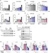
Fig. 8. hCitH3-mAb preserves macrophage integrity and mitigates CitH3-induced cytokine storm.
a LDH release from supernatants of BMDMs treated with increasing doses of CitH3 or unmodified H3 peptides for 24 h at 37 °C, showing CitH3-induced cytotoxicity (n = per group). b Cell viability, assessed using the CCK8 assay, demonstrated significant reduction following CitH3 peptide treatment compared to the H3 peptide (n = 5 per group). c BMDMs treated with 50 µg/mL CitH3 peptide and increasing doses of hCitH3-mAb or human IgG. LDH release was measured to evaluate cytotoxicity (n = per group).d, Viability of BMDMs treated as in (c) was measured using the CCK8 assay. hCitH3-mAb significantly preserved cell viability compared to control IgG (n = per group). e Immunoblot analysis of CitH3 in BMDMs treated with 15 μg/mL CitH3 or H3 peptides. Cells were washed three times prior to collection. And given that the CitH3 peptide is only ~30 amino acids in length, the bands observed at ~17 kDa are interpreted as endogenous cellular CitH3 protein detected at various time points. Similar results from three independent replicates. f Western blot analysis of citrullinated proteins in BMDMs from WT, Pad2⁻/⁻, and Pad2/4⁻/⁻ mice following 1 h treatment with 15 μg/mL CitH3 peptide. CitH3-induced citrullination was PAD2-dependent. Similar results from four independent replicates. g Quantification of IL-6, IL-1β, TNFα, IFN-α, and IFN-β levels in the supernatants of BMDMs treated with 25 μg/mL CitH3 peptide for 24 h (n = 6 per group). hCitH3-mAb effectively reduced CitH3-induced cytokine elevation in a dose-dependent manner, whereas control IgG had no effect. Data are presented as mean ± SD. Statistical analysis: One-way ANOVA followed by Dunnett’s multiple comparisons test was applied throughout. Comparisons were made to the ‘0’ group in (a–d), and the CitH3-treated group in (g), as indicated. Significance thresholds: *p < 0.05, **p < 0.01, ***p < 0.001, ****p < 0.0001.Source data are provided as a Source Data file.

References

    1. Singer, M. et al. The Third International Consensus Definitions for Sepsis and Septic Shock (Sepsis-3). JAMA315, 801–810 (2016). - PMC - PubMed
    1. Angus, D. C. & van der Poll, T. Severe sepsis and septic shock. N. Engl. J. Med369, 2063 (2013). - PubMed
    1. Marshall, J. C. Inflammation, coagulopathy, and the pathogenesis of multiple organ dysfunction syndrome. Crit. Care Med29, S99–S106 (2001). - PubMed
    1. Prescott, H. C. & Angus, D. C. Enhancing Recovery From Sepsis: A Review. JAMA319, 62–75 (2018). - PMC - PubMed
    1. Hotchkiss, R. S., Monneret, G. & Payen, D. Sepsis-induced immunosuppression: from cellular dysfunctions to immunotherapy. Nat. Rev. Immunol.13, 862–874 (2013). - PMC - PubMed

MeSH terms