. 2025 Aug 13;30(1):748.
doi: 10.1186/s40001-025-03012-5.

FGFR2 identified as a NETs-associated biomarker and therapeutic target in diabetic foot ulcers

Affiliations

FGFR2 identified as a NETs-associated biomarker and therapeutic target in diabetic foot ulcers

Linrui Dai et al. Eur J Med Res. .

Abstract

Background: Diabetic foot ulcer (DFU) is characterized by impaired wound healing and chronic inflammation, partly driven by the excessive formation of neutrophil extracellular traps (NETs). However, the molecular mediators linking NETs to failed tissue regeneration remain poorly understood. This study aimed to identify and validate novel NETs-associated biomarkers in DFU using an integrative bioinformatics and machine learning approach.

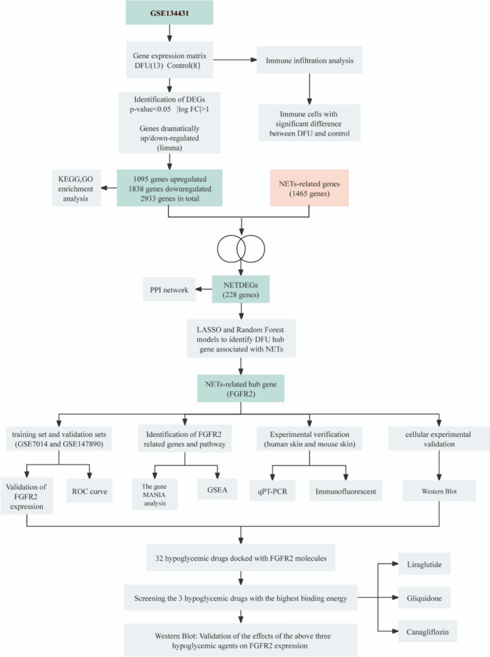
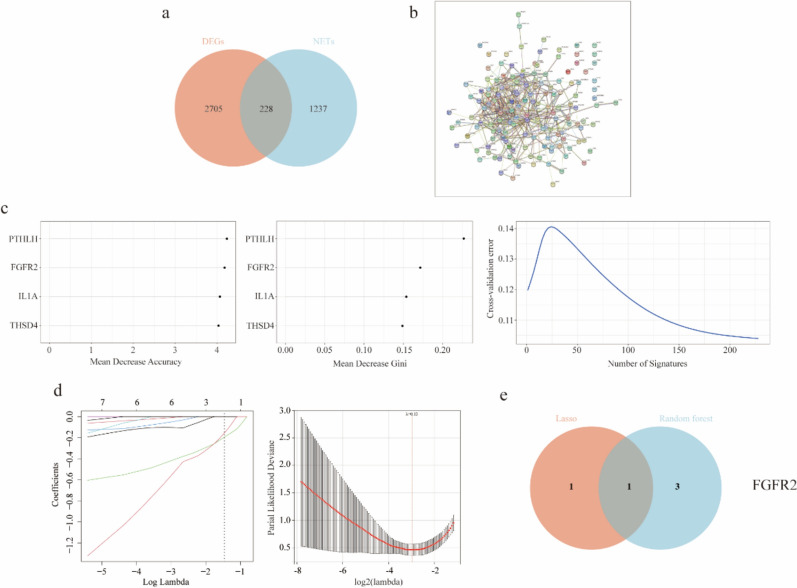
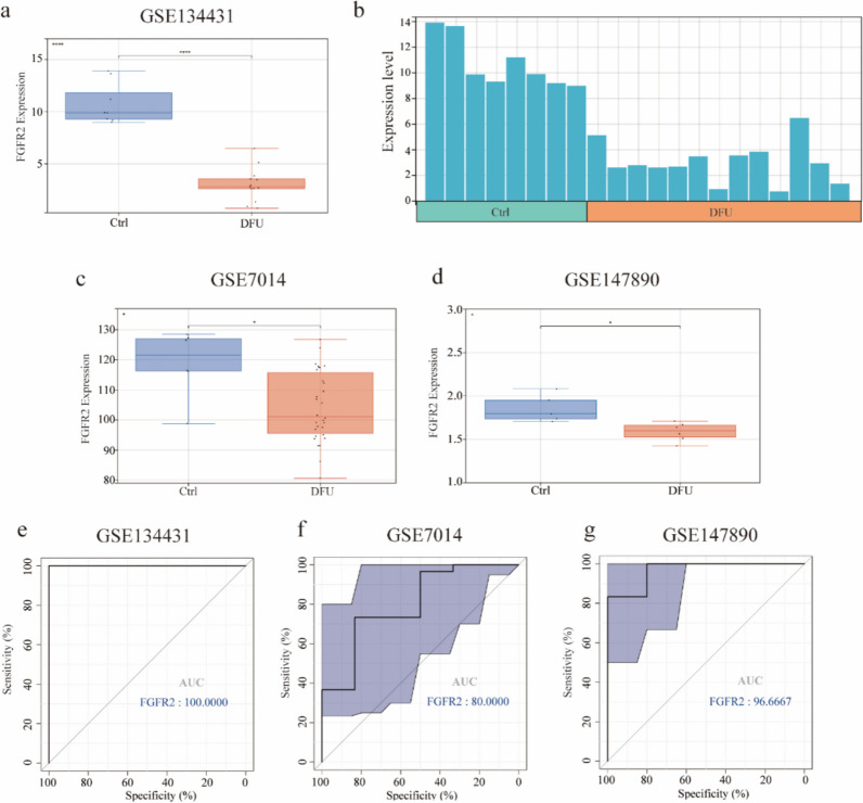
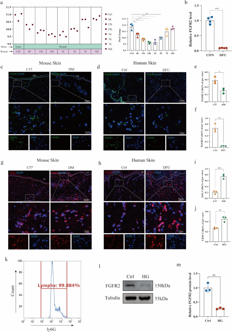
Methods: Differentially expressed genes (DEGs) were identified from the GEO dataset GSE134431. These DEGs were intersected with a NETs-related gene set to identify NETs-associated DEGs (NETDEGs). LASSO logistic regression and Random Forest algorithms were applied to the NETDEGs to select key feature genes. The top candidate, Fibroblast Growth Factor Receptor 2 (FGFR2), was validated in two independent datasets (GSE7014 and GSE147890). Its expression was further confirmed in human DFU tissues and diabetic mouse models using qPCR and immunohistochemistry. The effect of a high-glucose environment on FGFR2 expression in neutrophils was assessed in vitro. Finally, molecular docking technique was used to screen for existing drugs targeting FGFR2, with top candidates validated at the cellular level.

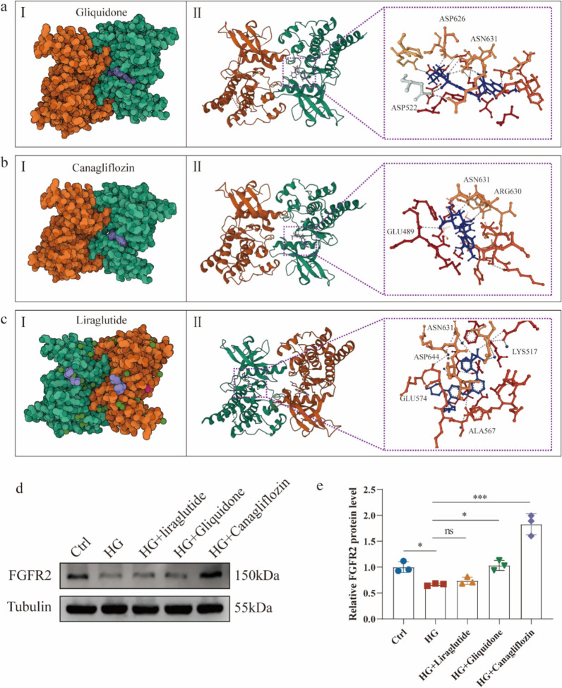
Results: Our analysis identified FGFR2 as a key downregulated gene at the intersection of DFU pathology and NETs-related pathways. FGFR2 expression was significantly reduced in DFU tissues across all datasets and in our experimental models, where its downregulation correlated with increased NETs accumulation. FGFR2 demonstrated strong diagnostic potential, with an AUC of 1.00 in the training set. In vitro, high glucose conditions suppressed FGFR2 expression in neutrophils. From 32 glucose-lowering drugs, Canagliflozin and Gliquidone were found to significantly upregulate FGFR2 protein expression in neutrophils, suggesting a potential modulatory effect.

Conclusions: FGFR2 is a promising potential biomarker associated with NET-driven inflammation in DFU. Its downregulation in the diabetic wound microenvironment and its modulation by existing pharmacological agents suggest that targeting the FGFR2 pathway may be a viable future therapeutic strategy for improving DFU healing outcomes. Further preclinical validation is warranted.

Keywords: Bioinformatics; Biomarker; DFU; FGFR2; NETs; Therapeutic Target.


Conflict of interest statement

Declarations. Ethics approval and consent to participate: The study was approved by the Ethics Committee of the Affiliated Central Hospital of Chongqing University with the approval number: 2023 Lun Audit No. (32). All participants gave informed consent. This study was guided by the Animal Welfare Ethics Committee of Chongqing University, and the ethical review number for animal experiments was CQU-IACUC-RE-202401-003. Consent for publication: All authors consent to the publication of this study. Competing interests: The authors declare no competing interests.

Figures

Fig. 1
Fig. 1
Research framework
Fig. 2
Fig. 2
Analysis of dataset variance and functional enrichment. a Heatmap of differential analysis between DFU group and control group; b Volcano plot of differential analysis between DFU group and control group; c KEGG pathway analysis of the intersection of genes. Different colors represent various significant pathways and related enriched genes; df GO analysis of the intersection of genes, including biological process, cellular component, and molecular function, respectively. The y-axis represents different GO terms, the x-axis represents gene ratio enriched in relative GO terms, the circle size refers to gene numbers, and the color represents the p-value
Fig. 3
Fig. 3
Immune cell infiltration analysis. a Stacked bar chart of the immune cell; b The correlation matrix of immune cell proportions; c: The box plot of the immune cell proportions
Fig. 4
Fig. 4
Screening hub genes using machine learning methods. a Venn plots of genes screened by DEGs and NETs-related gene datasets; b The PPI network of NETs-associated DEGs; c The Random Forest algorithm shows the DEGs associated with NETs and ranks them according to importance score genes; d Biomarker screening in the Lasso model; e The Venn diagram shows that a total of 1 gene was identified by the above two algorithms, FGFR2
Fig. 5
Fig. 5
Differences in FGFR2 genes between training and validation sets. a Differences in FGFR2 gene expression between DFU and control groups within the training set; b Expression profiles of the epidermal growth factor receptor 2 genes in each sample of the training set; c Differences in FGFR2 gene expression between DFU and control groups in the validation set GSE7014; d Differences in FGFR2 gene expression between DFU and control groups in the validation set GSE147890; e: ROC curve for FGFR2 in GSE134431; f ROC curve of FGFR2 in GSE7014; g ROC curve of FGFR2 in GSE147890
Fig. 6
Fig. 6
Analysis of FGFR2-related genes and functions. a Co-expressed genes of FGFR2 were analyzed by GeneMANIA; b Enrichment maps from genomic GESA enrichment analysis in DFU
Fig. 7
Fig. 7
FGFR2 expression and NETs level verification. a The relative expression levels of FGFR2 mRNA, during the course of cutaneous wound healing (days 0, 1, 3, 5, 7, and 10), were determined by extracting data from an existing RNA-seq gene expression dataset (GSE23006). Each time-point included three unique samples (N = 3); b The expression of FGFR2 in DFUs and the controls was analyzed by qPCR (N = 3); c IF of FGFR2 (green) in skin tissues of mice with diabetic wounds (N = 3); d IF of FGFR2 (green) in skin tissues of patients with diabetic wounds (N = 3); e, f Quantification of FGFR2; g IF of ly6G (red) in skin tissues of mice with diabetic wounds (N = 3); h IF of CD15 (red) in skin tissues of patients with diabetic wounds (N = 3); i, j Quantification of ly6G and CD15; k Flow cytometry validation of ly6G-labeled neutrophil ratio (N = 3); l Western blot validates the effect of a high glucose model on neutrophil FGFR2 expression (N = 3); m Western blot statistics. Experiments were performed at least in triplicate, and the results are shown as the mean ± s.d. *P < 0.05, **P < 0.01, ***P < 0.001
Fig. 8
Fig. 8
Screening Glucose-Lowering Drugs Targeting FGFR2. a binding mode of Gliquidone to FGFR2; b binding mode of Canagliflozin to FGFR2; c binding mode of Liraglutide to FGFR2 (I: Cartoon representation of the crystal structure of the small molecule compounds as well as the superposition of the target; II: 3D visualization of the binding pocket, generated with PyMOL software); d Western Blot validation of the effects of three hypoglycemic drugs on FGFR2 expression levels under hyperglycemic conditions; e Western Blot statistics from three independent experiments. Data are shown as mean ± s.d. *P < 0.05, ***P < 0.001

Similar articles

References

    1. Armstrong DG, Tan TW, Boulton AJM, Bus SA. Diabetic foot ulcers: A review. JAMA. 2023;330(1):62-75. - PMC - PubMed
    1. Yang M, Deng B, Hao W, Jiang X, Chen Y, Wang M, et al. Platelet concentrates in diabetic foot ulcers: a comparative review of PRP, PRF, and CGF with case insights. Regen Ther. 2025;28:625-32. - PMC - PubMed
    1. Deng L, Du C, Song P, Chen T, Rui S, Armstrong DG, et al. The role of oxidative stress and antioxidants in diabetic wound healing. Oxid Med Cell Longev. 2021;2021:8852759. - PMC - PubMed
    1. Ndosi M, Wright-Hughes A, Brown S, Backhouse M, Lipsky BA, Bhogal M, et al. Prognosis of the infected diabetic foot ulcer: a 12-month prospective observational study. Diabet Med. 2018;35(1):78–88. - PMC - PubMed
    1. Chen T, Song P, He M, Rui S, Duan X, Ma Y, et al. Sphingosine-1-phosphate derived from PRP-Exos promotes angiogenesis in diabetic wound healing via the S1PR1/AKT/FN1 signalling pathway. Burns Trauma. 2023;11:tkad003. - PMC - PubMed

Substances

LinkOut - more resources