

Development of in-house software to process real-time cine magnetic resonance images acquired during 1.5 T MR-guided radiation therapy
- PMID: 40797010
- PMCID: PMC12343943
- DOI: 10.1038/s41598-025-15107-4
Development of in-house software to process real-time cine magnetic resonance images acquired during 1.5 T MR-guided radiation therapy
Abstract
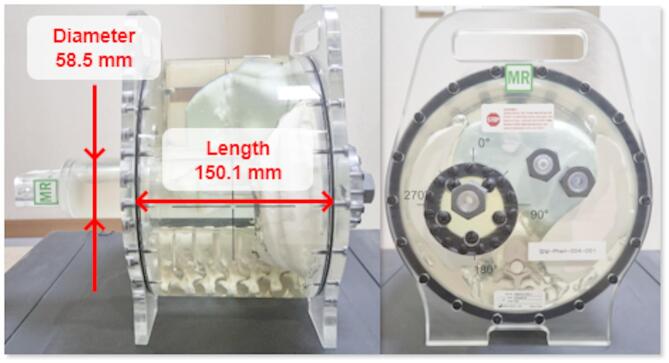
This study aimed to develop and publicly release an in-house software package that converts the binary file format of 2D cine magnetic resonance (MR) images acquired through the Treatment Session Manager (TSM) of MOSAIQ on the Elekta Unity (Elekta AB, Stockholm, Sweden) into standard readable data formats. The software was developed using MATLAB (MathWorks, Natick, MA, USA) and includes an automatic image-sorting algorithm to classify the images into coronal, sagittal, and axial planes. To verify the geometric accuracy of the converted images, they were acquired from an MRgRT motion management QA phantom, both with and without motion. For the converted images without motion, the geometric size of the phantom was measured and compared with the known values. For the images acquired with motion, the magnitudes measured using the converted cine MR images were compared with the artificially introduced motions. The results showed that the 2D cine MR images in the binary file format were successfully converted into the metadata/DICOM format and accurately classified into different planes. The accuracies of the geometric size and motion in the converted 2D cine MR images were greater than 99.6% and 94.2%, respectively. This software is expected to be useful for 1.5 T MR-linac users in analyzing internal organ motion during radiation treatment.
Keywords: 2D cine MR image; MATLAB; MR guided radiotherapy; MR-linac.
© 2025. The Author(s).
Conflict of interest statement
Declarations. Competing interests: The authors declare no competing interests. Conflict of interest: The authors declare that they have no known competing financial interests or personal relationships that could have appeared to influence the work reported in this paper.
Figures


References
-
- Mcdonald, B. A. et al. Initial feasibility and clinical implementation of daily MR-Guided adaptive head and neck cancer radiation therapy on a 1.5T MR-Linac system: prospective R-IDEAL 2a/2b systematic clinical evaluation of technical innovation. Int. J. Radiat. Oncol. Biol. Phys.109, 1606–1618. 10.1016/j.ijrobp.2020.12.015 (2021). - DOI - PMC - PubMed
MeSH terms
Grants and funding
LinkOut - more resources
Full Text Sources
