This is a preprint.
Loss of Ag85A disrupts plasma membrane domains and promotes free mycolic acid accumulation in mycobacteria
- PMID: 40799537
- PMCID: PMC12340856
- DOI: 10.1101/2025.08.05.668640
Loss of Ag85A disrupts plasma membrane domains and promotes free mycolic acid accumulation in mycobacteria
Abstract
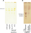
The mycomembrane of mycobacteria, composed primarily of long-chain mycolic acids, is critical for cell survival, structural integrity, and resistance to environmental stress, yet its underlying synthesis mechanisms remain incompletely understood. This study investigates the role of Ag85A, a key enzyme in mycomembrane synthesis, in regulating plasma membrane domains and cell envelope organization in Mycobacterium smegmatis. Using ΔAg85A deletion mutants, we combined microscopy, biochemical assays, thin-layer chromatography, and lipid analysis to evaluate changes in membrane structure, chemical accumulation, and lipid composition. Ag85A deletion leads to altered plasma membrane domain organization, increased chemical accumulation, changes in cell envelope lipid composition. Unexpectedly, lipid analysis revealed accumulation-not depletion-of mycolic acids in the mutant, suggesting that increased permeability is not directly due to mycolic acid loss. These findings highlight a novel link between mycomembrane composition and plasma membrane domain stability. Our study not only advances understanding of mycobacterial cell envelope architecture but also identifies potential targets for enhancing drug penetration in resistant mycobacterial infections.
Keywords: Antigen 85A; Membrane domain; Membrane permeability; Mycobacteria; Mycolic acids.
Figures


Similar articles
-
Disulfide bonds are critical for stabilizing cell division, cell envelope biogenesis, and antibiotic resistance proteins in mycobacteria.mBio. 2025 Sep 10;16(9):e0108325. doi: 10.1128/mbio.01083-25. Epub 2025 Jul 31. mBio. 2025. PMID: 40741763 Free PMC article.
-
A conditional mutant of the fatty acid synthase unveils unexpected cross talks in mycobacterial lipid metabolism.Open Biol. 2017 Feb;7(2):160277. doi: 10.1098/rsob.160277. Open Biol. 2017. PMID: 28228470 Free PMC article.
-
Role of long-chain acyl-CoAs in the regulation of mycolic acid biosynthesis in mycobacteria.Open Biol. 2017 Jul;7(7):170087. doi: 10.1098/rsob.170087. Open Biol. 2017. PMID: 28724694 Free PMC article.
-
Antibiotic treatment for non-tuberculous mycobacteria lung infection in people with cystic fibrosis.Cochrane Database Syst Rev. 2025 Mar 27;3(3):CD016039. doi: 10.1002/14651858.CD016039. Cochrane Database Syst Rev. 2025. PMID: 40145528
-
Decoding the Penicillin-Binding Proteins with Activity-Based Probes.Acc Chem Res. 2025 Jun 3;58(11):1754-1763. doi: 10.1021/acs.accounts.5c00113. Epub 2025 May 21. Acc Chem Res. 2025. PMID: 40396497 Review.
References
-
- Carranza C, Chavez-Galan L. 2019. Several routes to the same destination: inhibition of phagosome-lysosome fusion by Mycobacterium tuberculosis. Am J Med Sci 357:184–194. - PubMed
-
- Dulberger CL, Rubin EJ, Boutte CC. 2020. The mycobacterial cell envelope — a moving target. 1. Nat Rev Microbiol 18:47–59. - PubMed
Publication types
Grants and funding
LinkOut - more resources
Full Text Sources
