

This is a preprint.
Miniaturized widefield microscope for high speed in vivo voltage imaging
- PMID: 40799544
- PMCID: PMC12340831
- DOI: 10.1101/2025.08.04.668551
Miniaturized widefield microscope for high speed in vivo voltage imaging
Update in
-
Miniaturized widefield microscope for high speed in vivo voltage imaging.Biomed Opt Express. 2025 Dec 2;17(1):1-11. doi: 10.1364/BOE.576516. eCollection 2026 Jan 1. Biomed Opt Express. 2025. PMID: 41532107 Free PMC article.
Abstract
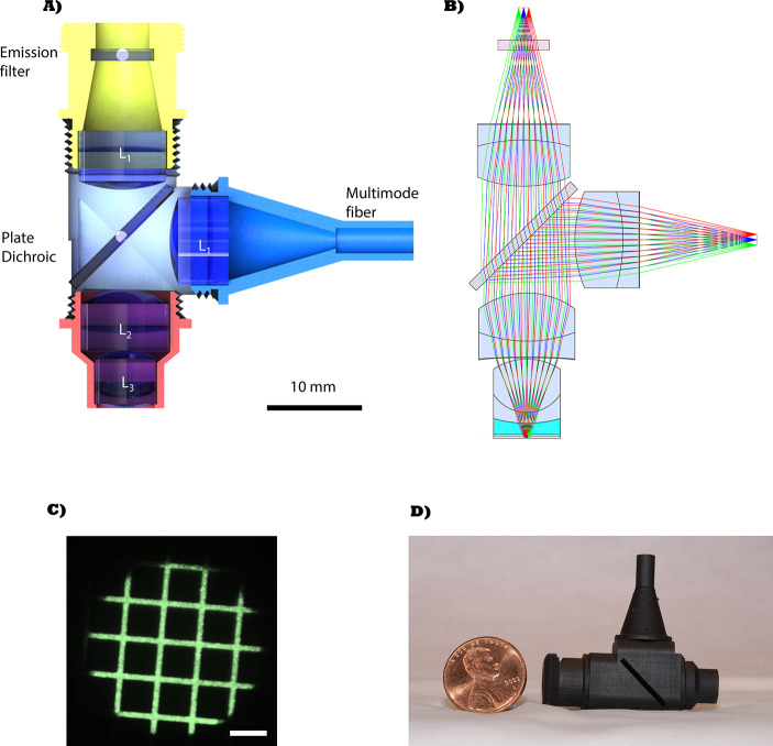
Functional imaging in freely moving animals with genetically encoded voltage indicators (GEVIs) will open new capabilities for neuroscientists to study the behavioral relevance of neural activity with high spatial and temporal precision. However, miniaturization of an imaging system with sufficient collection efficiency to resolve the small changes in fluorescence yield from voltage spikes, as well as development of efficient image sensors that are sufficiently fast to capture them, has proven challenging. We present a miniaturized microscope designed for voltage imaging, with a numerical aperture of 0.6, 250 μm field of view and 1.3 mm working distance that weighs 16.4 g. We show it is capable of imaging in vivo voltage spikes from Voltron2 with a spike peak-to-noise ratio >3 at a framerate of 530 Hz.
Conflict of interest statement
Disclosures. The authors declare no conflict of interest.
Figures


References
-
- Bai T., Zhan L., Zhang N., et al. , “Learning-prolonged maintenance of stimulus information in CA1 and subiculum during trace fear conditioning,” Cell Reports 42 (2023). - PubMed
-
- Simoes de Souza F. M., Williamson R., McCullough C., et al. , “Miniscope Recording Calcium Signals at Hippocampus of Mice Navigating an Odor Plume,” J. visualized experiments : JoVE (2024). - PubMed
Publication types
Grants and funding
LinkOut - more resources
Full Text Sources