[Preprint]. 2025 Aug 4:2025.08.04.668444.
doi: 10.1101/2025.08.04.668444.

Genetic mechanisms of resistance to targeted KRAS inhibition

Affiliations

Genetic mechanisms of resistance to targeted KRAS inhibition

Bianca J Diaz et al. bioRxiv. .

Abstract

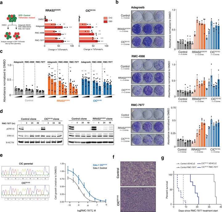
KRAS mutations are among the most prevalent oncogenic drivers in non-small cell lung cancer (NSCLC), yet the mechanisms of therapeutic resistance to KRAS inhibitors in these cancers remains poorly understood. Here, we deploy high-throughput CRISPR base editing screens to systematically map resistance mutations to three mechanistically distinct KRAS-targeted therapies, including KRAS-G12C(OFF) inhibitor (adagrasib), RAS(ON) G12C-selective tri-complex inhibitor (RMC-4998), and RAS(ON) multi-selective tri-complex inhibitor (RMC-7977). Using both a saturation Kras tiling approach and cancer-associated mutation library, we identify common and compound-selective second-site resistance mutations in Kras, as well as gain-of-function and loss-of-function variants across cancer-associated genes that rewire signaling networks in a context-dependent manner. Notably, we identify a recurrent missense mutation in capicua (Cic), that promotes resistance to RMC-7977 in vitro and in vivo. Moreover, we show that targeting NFκB signaling in CIC-mutant cells can resensitize them to RAS pathway inhibition and overcome resistance.


Conflict of interest statement

Conflict of Interest Statement LED is a consultant for and holds equity in Mirimus Inc., unrelated to this work. LED is a consultant for and has received grant funding from Revolution Medicines, Inc. related to this work. AT, NAV, AK, MPL, IA, and MS are employees and stockholders of Revolution Medicines, Inc.

Figures

Figure 1.
Figure 1.. A new computational pipeline to analyze BE tiling libraries.
a, Schematic methodology to derive 2D lung tumor lines. b, Measurement of cytosine base editing efficiency with GO reporter by GFP expression using flow cytometry. c, Schematic of the Kras-TILE library cloned into our library sensor vector and transduced into cells expressing CBE or ABE. d, Comparison of fraction of reads output from merged fastqs from D0 samples of CBE and ABE cells (n = 3) transduced with Kras-TILE either using exact matching or probabilistic matching to the whitelist. e, Correlation of BE editing activity detected at sensor to sgRNA self-editing at D0 across cells transduced with FNLS (NGG), FNLS-NG, and FNLS-SpRY and ABE8e-SpRY. f, Reads were pulled from individual sgRNAs with high BE activity in the Kras-TILE library and analyzed with CRISPResso2 at the sgRNA and sensor regions. C>T (orange) and A>G (blue) transitions were measured across all potential nucleotides. g, Schematic describing BEquant pipeline.
Figure 2.
Figure 2.. KRAS BE tiling identifies unique and shared hotspot resistance mutations to adagrasib and RMC-4998.
a, Percent BE at the target base (cytosine or adenine in the 5th position of sgRNA) across the Kras transcript (x axis). Dots are colored by the potential outcome of the predicted mutation. b, Schematic of Kras-TILE screen describing dosing and samples for NGS (n = 3 transduction replicates). c, Manhattan plot demonstrating the median log2 fold change of each sgRNA calculated with MAGeCK (y axis) across the Kras transcript (x axis) at each timepoint across Adagrasib and RMC-4998 treated samples (n = 3). d, Schematic of Kras WT and LSL-KrasG12C sequence depicting Kras-TILE sgRNAs from Adagrasib and RMC-4998 treated samples. e, Line graphs demonstrating changes in log2 fold change calculated with MAGeCK (y axis) of the G122R and H951F sgRNAs over time (x axis). Lines are colored by drug treatment.
Figure 3.
Figure 3.. KRAS BE tiling identifies unique and shared hotspot resistance mutations to RMC-7977
a. Manhattan plot demonstrating the median log2 fold change of each sgRNA calculated with MAGeCK (y axis) across the Kras transcript (x axis) at each timepoint in RMC-7977 treated samples (n = 3). b. Line graphs demonstrating changes in log2 fold change with MAGeCK (y axis) of the E633R and Y712R sgRNAs over time (x axis). Lines are colored by drug treatment. c. Stacked bar chart showing the frequency of specific alleles determined by amplification of the exon and NGS of the endogenous locus of Kras from the screen samples treated with DMSO or RMC-7977 at D20 for CBE (left n= 3) and ABE (right n=3). d. X-ray crystal structure of CYPA:RMC-7977:KRAS(GMPPNP)G12C (PDB:8TBK) shows that the SWII region of KRAS forms part of the protein-protein interface of the tri-complex. E63 is proximal to both RMC-7977 and CYPA residues W121 and K125 while Y71 forms a number of hydrophobic interactions within KRAS.
Figure 4.
Figure 4.. MAPK and inflammatory regulators drive resistance to KRAS inhibitors.
a, MBES screen timeline schematic describing dosing and samples for NGS (n = 5–6 transduction replicates). b, Bubbleplot comparing sgRNA log2 fold change (y axis) and mean sensor base editing (x axis) at D20 in DMSO control treated samples. Colors represent predicted variant and size of dot represents mean BE% at D0. c, Volcano plots of sgRNAs that are significantly enriched in blue and significantly depleted in orange from Adagrasib, RMC-4998, and RMC-7977 treated samples compared to DMSO at D20 (left to right). d, (left) OncoKB annotation of each enriched sgRNA at D20 and its predicted mutation outcome and its predicted oncogenic effect. (right) Heatmap of D20 enriched sgRNAs as rows and each column representing a replicate and binned by its associated treatment condition. Each box is colored by its log2 fold change score calculated with MAGeCK. e, Pie charts (left) of predicted functional outcomes of sgRNAs in the MBES library and (right) of predicted functional outcomes of sgRNAs from the enriched hits at D20.
Figure 5.
Figure 5.. CIC mutations promote resistance to multi-selective RAS inhibition in mouse and human cells and in vivo in NSCLC.
a, (left) Schematic of the competition assay methodology. KCP4, 5, and 6 cells are individually transduced with a control sgRNA in a GFP expressing guide plasmid and a mutant sgRNA in a TdTomato expressing guide plasmid and selected for 7 days with blasticidin. Selected cells are mixed in a 80:20 control to mutant cell ratio and either treated with DMSO, Adagrasib (250nM), RMC-4998 (250nM), and RMC-7977 (30nM). Samples were analyzed for the GFP and TdTomato expression at D3 (initial timepoint) and D10 and D20. (right) Quantification of change in positive TdTomato population % from D3 at D10 and D20 from each treatment group in Rras2G23D/N and CicR215Q mutant cells (n = 3 biological replicates and performed 2 independent times). Two-way ANOVA (analysis of variance); *P < 0.05, **P < 0.01 ***P < 0.0001. b, (left) Representative images of endpoint of colony formation assay of 3 independently derived singlcell clones after treatment with Adagrasib (250nM), RMC-4998 (250nM), and RMC-7977 (30nM). (right) Quantification of Giemsa stain as measured by absorbance normalized to DMSO treated cells for 6 independently derived clones per mutant in KCP4 and KCP5 cells. c, Summary quantification of all colony formation assay experiments performed. Each bar is an increasing dose of Adagrasib (100nM, 250nM, and 750nM), RMC-4998 (100nM, 250nM, and 750nM), and RMC-7977 (5nM, 30nM, and 100nM). Each dot represents the mean absorbance value of an individual single cell clone performed 3 independent times (n = 2 independent cell lines and 3 independent single cell clones). Two-way ANOVA (analysis of variance); *P < 0.05, **P < 0.01 ***P < 0.0001. d, Western blotting of control and mutant single cell clones from KCP4 cells after treatment with RMC-7977 30nM and protein samples collected at 4, 24, and 48 hours where expression of phosphorylated ERK is assessed. e, (left) Sanger sequencing traces of CIC R215 sgRNA locus from Calu-1 control and Calu-1 CICR215Q single cell clone. (right) RMC-7977 dose response assay comparing cell viability at D6. Data represents 3 independently derived single cell clones and performed 2 separate times. Two-way ANOVA (analysis of variance); ***P < 0.0001. f, Representative H&E images from vehicle treated control or CicR15Q mice (n = 3–5/group) g, Survival analysis of mice transplanted with control or CicR215Q cells (n = 3–5/group). **P < 0.01. P values were calculated using the log-rank test.
Figure 6:
Figure 6:. Targeting NFκB synergizes with RMC-7977 to suppress outgrowth of CIC mutant cells.
a, Heatmap showing average Log2 normalized expression of differentially expressed genes from KRAS targets, LeBlanc CIC targets, and NFkB targets gene sets across DMSO and RMC-7977 30nm treated samples (n = 6 independently derived single cell clones from KCP4 and KCP5 cells). b, Summary of the gene sets significantly enriched in CICR215Q mutant samples compared to control, NF2x225_splice, and RRAS2G23D/N mutant samples treated with RMC-7977 30nM. Size and color of dot represents NES. Shading of dot represents significance (n = 6 independently derived single cell clones from KCP4 and KCP5 cells). c, Dotplot comparing CIC target pathway enrichment z scores from two published datasets Okimoto CIC targets (x axis) and LeBlanc CIC targets (y axis). d, (left) Representative image of colony formation assay experiment of endpoint of control and CICR215Q cells treated with RMC-7977 30nM, IKK-16 1uM, or the combination. (right) Quantification of Giemsa stain absorbance value normalized to DMSO. (n = 3 independently derived single cell clones from KCP5 cells and performed 2 separate times). Two-way ANOVA (analysis of variance); *P < 0.05 and ***P < 0.0001. e, Control and CICR215Q mutant cells were treated with indicated concentration of DMSO, RMC-7977, and IKK-16 for 6 days and synergy was calculated based on cell viability at different dose pairs. Data are mean of 3 biological replicates. f, Summary of Max synergy scores across control and CICR215Q mutant samples.

References

    1. Society, A. C. Facts & Figures 2025, <https://www.cancer.org/cancer/types/lung-cancer/about/key-statistics.html> (2025).
    1. Canon J. et al. The clinical KRAS(G12C) inhibitor AMG 510 drives anti-tumour immunity. Nature 575, 217–223 (2019). 10.1038/s41586-019-1694-1 - DOI - PubMed
    1. Skoulidis F. et al. Sotorasib for Lung Cancers with KRAS p.G12C Mutation. New England Journal of Medicine 384 (2021-June-24). 10.1056/NEJMoa2103695 - DOI - PMC - PubMed
    1. Christensen J. G. et al. The KRASG12C Inhibitor, MRTX849, Provides Insight Toward Therapeutic Susceptibility of KRAS Mutant Cancers in Mouse Models and Patients. Cancer Discovery, CD-19–1167 (2019). 10.1158/2159-8290.Cd-19-1167 - DOI - PMC - PubMed
    1. Jänne P. A. et al. Adagrasib in Non–Small-Cell Lung Cancer Harboring a KRASG12C Mutation. New England Journal of Medicine 387 (2022-July-14). 10.1056/NEJMoa2204619 - DOI - PubMed

Publication types

LinkOut - more resources