

Dual-encoded magnetization transfer and diffusion imaging and its application to tract-specific microstructure mapping
- PMID: 40799701
- PMCID: PMC12007536
- DOI: 10.1162/imag_a_00019
Dual-encoded magnetization transfer and diffusion imaging and its application to tract-specific microstructure mapping
Abstract
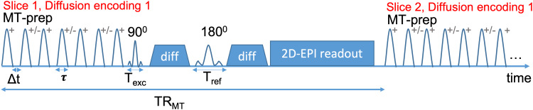
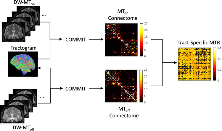
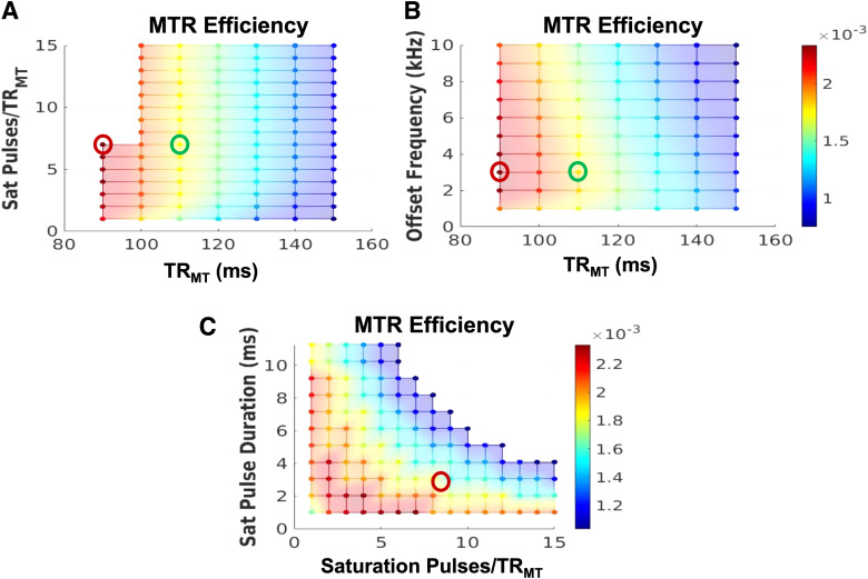
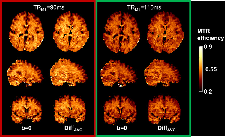
We present a novel dual-encoded magnetization transfer (MT) and diffusion-weighted sequence and demonstrate its potential to resolve distinct properties of white matter fiber tracts at the sub-voxel level. The sequence was designed and optimized for maximal MT ratio (MTR) efficiency. The resulting whole brain 2.6 mm isotropic protocol to measure tract-specific MTR has a scan time under 7 minutes. Ten healthy subjects were scanned twice to assess repeatability. Two different analysis methods were contrasted: a technique to extract tract-specific MTR using Convex Optimization Modeling for Microstructure Informed Tractography (COMMIT), a global optimization technique; and conventional MTR tractometry. The results demonstrate that the tract-specific method can reliably resolve the MT ratios of major white matter fiber pathways and is less affected by partial volume effects than conventional multi-modal tractometry. By reducing the contamination due to partial volume averaging of tracts, dual-encoded MT and diffusion may increase the sensitivity to microstructure alterations of specific tracts due to disease, aging, or learning, as well as lead to weighted structural connectomes with more anatomical specificity.
Keywords: connectome; diffusion; dual-encoding; magnetization transfer; microstructure; myelin.
© 2023 Massachusetts Institute of Technology.
Conflict of interest statement
None to declare.
Figures
Similar articles
-
Mapping the aggregate g-ratio of white matter tracts using multi-modal MRI.Imaging Neurosci (Camb). 2025 Jun 18;3:IMAG.a.49. doi: 10.1162/IMAG.a.49. eCollection 2025. Imaging Neurosci (Camb). 2025. PMID: 40800937 Free PMC article.
-
Prescription of Controlled Substances: Benefits and Risks.2025 Jul 6. In: StatPearls [Internet]. Treasure Island (FL): StatPearls Publishing; 2025 Jan–. 2025 Jul 6. In: StatPearls [Internet]. Treasure Island (FL): StatPearls Publishing; 2025 Jan–. PMID: 30726003 Free Books & Documents.
-
Translating state-of-the-art spinal cord MRI techniques to clinical use: A systematic review of clinical studies utilizing DTI, MT, MWF, MRS, and fMRI.Neuroimage Clin. 2015 Dec 4;10:192-238. doi: 10.1016/j.nicl.2015.11.019. eCollection 2016. Neuroimage Clin. 2015. PMID: 26862478 Free PMC article.
-
MarkVCID cerebral small vessel consortium: II. Neuroimaging protocols.Alzheimers Dement. 2021 Apr;17(4):716-725. doi: 10.1002/alz.12216. Epub 2021 Jan 21. Alzheimers Dement. 2021. PMID: 33480157 Free PMC article.
-
Management of urinary stones by experts in stone disease (ESD 2025).Arch Ital Urol Androl. 2025 Jun 30;97(2):14085. doi: 10.4081/aiua.2025.14085. Epub 2025 Jun 30. Arch Ital Urol Androl. 2025. PMID: 40583613 Review.
References
-
- Andersson, J. L., Skare, S., & Ashburner, J. (2003). How to correct susceptibility distortions in spin-echo echo-planar images: Application to diffusion tensor imaging. NeuroImage, 20(2), 870–888. https://doi.org/10.1016/S1053-8119(03)00336-7Get rights and content - PubMed
-
- Barakovic, M., Girard, G., Schiavi, S., Romascano, D., Descoteaux, M., Granziera, C., Jones, D. K., Innocenti, G. M., Thiran, J.-P., & Daducci, A. (2021). Bundle-specific axon diameter index as a new contrast to differentiate white matter tracts. Front Neurosci, 15, 646034. 10.3389/fnins.2021.646034 - DOI - PMC - PubMed
-
- Barakovic, M., Tax, C. M. W., Rudrapatna, U., Chamberland, M., Rafael-Patino, J., Granziera, C., Thiran, J. P., Daducci, A., Canales-Rodríguez, E. J., & Jones, D. K. (2021). Resolving bundle-specific intra-axonal T2 values within a voxel using diffusion-relaxation tract-based estimation. NeuroImage, 227, 117617. 10.1016/j.neuroimage.2020.117617 - DOI - PMC - PubMed
-
- Battiston, M., Schneider, T., Grussu, F., Yiannakas, M. C., Prados, F., De Angelis, F., Gandini Wheeler-Kingshott, C. A. M., & Samson, R. S. (2019). Fast bound pool fraction mapping via steady-state magnetization transfer saturation using single-shot EPI. Magn Reson Med, 82(3), 1025–1040. 10.1002/mrm.27792 - DOI - PubMed
LinkOut - more resources
Full Text Sources