[Preprint]. 2025 Aug 4:rs.3.rs-7159889.
doi: 10.21203/rs.3.rs-7159889/v1.

Nuclear Envelope Membrane Protein 1 plays crucial and conserved roles in female meiosis

Affiliations

Nuclear Envelope Membrane Protein 1 plays crucial and conserved roles in female meiosis

Bilal Ahmad Hakim et al. Res Sq. .

Abstract

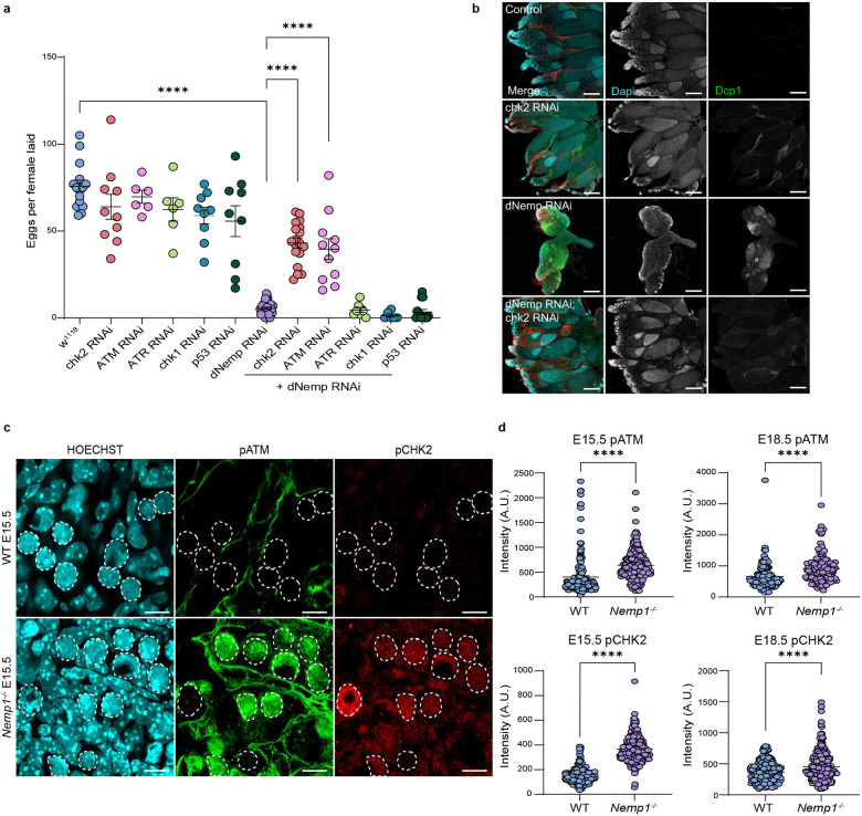
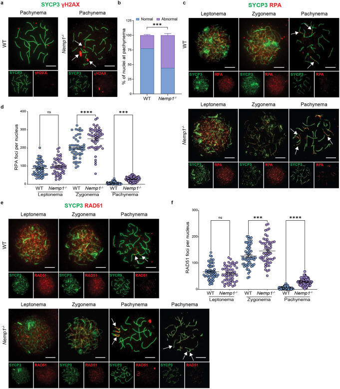
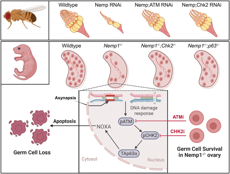
Female germ cells must preserve the integrity of their genome and generate genetic diversity via meiotic recombination. This challenging process, which occurs during fetal life, is error prone. Highly conserved checkpoint pathways detect errors in recombination and DNA damage, inducing the death of defective oocytes. Nuclear Envelope Membrane Protein (NEMP) homologs are highly conserved inner nuclear membrane proteins which are critical for fertility in flies, worms, fish and mice, and mechanically support the nuclear envelope. However, why NEMP homologs are required for fertility is still unclear. Using both Drosophila and mouse models, we establish here that loss of Nemp1 leads to activation of an ATM-CHK2 DNA damage pathway and results in massive loss of oocytes during fetal life. Chemical or genetic inactivation of the ATM-CHK2-p63 pathway reduces oocyte loss, demonstrating its importance upon loss of Nemp1. In the absence of Nemp1 meiotic progression is delayed and DNA damage is increased at zygonema and pachynema stages. Loss of Nemp1 also leads to defects in chromosome synapsis persisting through pachynema. We conclude that Nemp1 is needed for timely and precise execution of meiotic prophase and is crucial for accurate pairing and synapsis, oocyte developmental competence and survival.


Conflict of interest statement

Competing interests The authors declare that they have no competing interests. Additional Declarations: There is NO Competing Interest.

Figures

Figure 1.
Figure 1.. Loss of embryonic and postnatal oocytes in Nemp1−/− mice occurs due to apoptosis.
a) Follicle count showing a significant gradual loss of primordial follicles in Nemp1−/− ovaries at postnatal day 2 (P2), P4 and P10. *P < 0.05, ***P < 0.001, ****P < 0.0001 (Unpaired t-test). Each dot represents the number of primordial follicles per ovary. b) Representative images showing normal primordial follicles and fewer TUNEL-positive (apoptosis marker) primordial follicle oocytes in P4 WT control ovaries, while an increased number of TUNEL-positive primordial follicle oocytes in P4 Nemp1−/− ovaries (Scale Bars - 50 μm). c) Quantitative analysis of TUNEL-positive primordial follicles of P4 mice. *P < 0.05 (Unpaired t-test). Each dot represents number of TUNEL-positive primordial follicle oocytes per ovary. d) Activated caspase 3 (red), a marker of apoptosis, staining in WT vs Nemp1−/− P2 ovarian sections showing increased cleaved caspase 3 expression in Nemp1−/− sections (Scale Bars - 50 μm). e) Intensity analysis of cleaved caspase 3 in WT vs Nemp1−/− ovaries at P2. ***P < 0.001 (Unpaired t-test). f) Nemp1 staining (green) in P2 WT VASA (red) positive oocytes (Scale Bars - 5 μm). g) Immunofluorescence labeling of Nemp1 (green) in embryonic 17.5 (E17.5) meiotic prophase SCP3 (red) positive oocytes. DNA is stained with DRAQ5 (Magenta). h) Representative images of 3D and germ cell spots analysis of whole-mount E15.5 WT ovary stained with germ cell markers, DDX4 (green) and TRA98 (red) (Scale Bars - 50 μm). i) Germ cell count shows a normal oocyte number in Nemp1 mutant embryonic 15.5 (E15.5) ovaries but a significant progressive oocyte loss in E18.5 and post-natal day 0 ovaries. ****P < 0.0001, **P < 0.01 (Unpaired t-test). Each dot represents the number of oocytes per ovary.
Figure 2.
Figure 2.. Activation of ATM and CHK2 causes germ cell loss in dNemp flies and Nemp1−/− ovaries.
a) The number of eggs laid by 3-day-old females shows rescue of egg laying in dNemp RNAi flies by reduction of ATM and Chk2 (DNA double-strand break pathway) but not by depletion of ATR and Chk1 (DNA single-strand break pathway). ****P < 0.0001 (one-way ANOVA, Tukey’s multiple comparison test). b) Representative confocal images of ovaries dissected from 3-day-old females showing ovary size rescue by reduction of Chk2 in dNemp flies (Scale Bars - 200 μm). c) Immunostaining of pATM and pCHK2 in E15.5 WT and Nemp1−/− ovaries. Dashed circles show germ cells (Scale Bars - 10 μm). d) Fluorescence intensity analysis of pATM and pCHK2 in WT and Nemp1−/− E15.5 and E18.5 germ cells showed a significant increase in these cell cycle checkpoints in Nemp1−/− germ cells. ****P < 0.0001 (Mann-Whitney nonparametric test).
Figure 3.
Figure 3.. Inactivation of ATM, CHK2 and p63 pathway rescues primordial follicle loss in Nemp1−/− mice.
a) Representative immunofluorescence of solvent and ATM inhibitor treated P4 WT and Nemp1−/− ovaries after 2 days of in vitro culture (Scale Bars - 50 μm). b) Follicle quantification, c) γH2ax, and d) p63 fluorescence intensity of solvent and ATM inhibitor treated WT and Nemp1−/− ovaries showing ATM inhibitor rescue partial primordial follicle number and reduce DNA damage in Nemp1−/− primordial follicle oocytes. *P < 0.05, **P < 0.01, ****P < 0.0001 (Paired t-test and Wilcoxon nonparametric test). e) Quantification of relative mRNA expression of NOXA in P2 WT and Nemp1−/− ovaries by real-time PCR. **P < 0.01 (Unpaired t-test). f) Representative immunostaining of solvent and CHK2 inhibitor treated P4 WT and Nemp1−/− ovaries after 2 days of in vitro culture (Scale Bars - 50 μm). g) Follicle quantification, h) γH2ax, and i) p63 fluorescence intensity of solvent and CHK2 inhibitor treated WT and Nemp1−/− ovaries showing a partial rescue of primordial follicle number and p63 reduction in CHK2 inhibitor treated Nemp1−/− ovaries compared with solvent treated Nemp1−/− ovaries. *P < 0.05, ****P < 0.0001 (Paired t-test and Wilcoxon nonparametric test). j) Follicle count of 4-week-old control (WT), Chk2−/−, Nemp1−/−, Nemp1−/−;Chk2−/−, and Nemp1−/−;p63fl/fl ZP3+ mice ovaries show deletion of Chk2 and p63 rescues primordial follicle count in Nemp1−/− animals. ****P < 0.0001 (two-way ANOVA, Tukey’s multiple comparison test).
Figure 4.
Figure 4.. Nemp1 loss leads to persistent DNA damage in embryonic oocytes.
a) Representative images of WT and Nemp1−/− pachynema oocyte spreads at E17.5 showing persistent γH2AX (red) staining in Nemp1−/− oocytes indicating DNA Damage. Oocytes are immunostained for SYCP3 (green) and γH2AX (red) (Scale Bars - 10 μm). b) Proportion of oocytes with persistent γH2AX (abnormal) in WT and Nemp1−/− E17.5 pachynema spreads. ***P < 0.001 (Unpaired t-test). c) Representative images of WT and Nemp1−/− E17.5 oocytes stained with SYCP3 (green) and RPA (red) showing leptonema, zygonema, and pachynema stages (Scale Bars - 10 μm). d) Quantification of RPA foci from three meiotic stages. ****P < 0.0001, ***P < 0.001 (one-way ANOVA, Sidak’s multiple comparison test). e) Representative images of WT and Nemp1−/− E17.5 oocytes stained with SYCP3 (green) and RAD51 (red) showing leptonema, zygonema, and pachynema stages (Scale Bars - 10 μm). f) Quantification of RAD51 foci from three meiotic stages. ***P < 0.001, ****P < 0.0001 (one-way ANOVA, Sidak’s multiple comparison test).
Figure 5.
Figure 5.. Nemp1 loss leads to defects in synapsis and recombination.
a) Percentage of oocyte spreads in P0 WT and Nemp1−/− ovaries suggesting a delay in Meiosis I progression. **P < 0.01 (Unpaired t-test). b) Examples of fully synapsed and asynapsed oocytes, where the white arrow shows the site of asynapsis (Scale Bars - 10μm). c) Quantification of asynapsis. **P < 0.01 (Unpaired t-test). d) Representative images of WT and Nemp1−/− E17.5 oocytes stained with SYCP3 (green) and Hormad1 (red) showing leptonema, zygonema, pachynema, and diplonema stages (Scale Bars - 10μm). e) Percentage of nuclei at pachynema showing retention of Hormad1 staining in WT and Nemp1−/− oocytes at E17.5. ***P < 0.001 (Unpaired t-test). White arrows show Hormad1 retention in Nemp1−/− oocytes. f) Representative images of WT and Nemp1−/− E17.5 pachynema oocyte spread stained with SYCP3 (green) and MLH1 (red) (Scale Bars −10μm). g) Quantification of MLH1 foci at pachynema. ****P < 0.0001 (Unpaired t-test).
Figure 6.
Figure 6.. Graphical summary of Nemp1 deficiency triggered oocyte loss.
Loss of Nemp1 in oocytes leads to excessive DNA damage, which activates the ATM-mediated CHK2 checkpoint in both flies and mice. This activation further triggers p63, resulting in NOXA-dependent apoptosis. Inhibition of ATM or CHK2, as well as genetic inactivation of CHK2 or p63, prevents oocyte loss in Nemp1 mutant ovaries. This demonstrates that the loss of Nemp1 alone is sufficient to activate a conserved checkpoint pathway, emphasizing its crucial role in the nuclear envelope of germ cells for maintaining genome integrity.

Similar articles

References

    1. BAKER T. G. A QUANTITATIVE AND CYTOLOGICAL STUDY OF GERM CELLS IN HUMAN OVARIES. Proceedings of the Royal Society of London. Series B, Containing papers of a Biological character. Royal Society (Great Britain) 158, 417–433 (1963). - PubMed
    1. Kurilo L. F. Oogenesis in antenatal development in man. Hum Genet 57, 86–92 (1981). - PubMed
    1. Hunter N. Oocyte Quality Control: Causes, Mechanisms, and Consequences. (2017) doi: 10.1101/sqb.2017.82.035394. - DOI - PubMed
    1. Bolcun-Filas E., Rinaldi V. D., White M. E. & Schimenti J. C. Reversal of female infertility by Chk2 ablation reveals the oocyte DNA damage checkpoint pathway. Science (1979) 343, 533–536 (2014). - PMC - PubMed
    1. Rinaldi V. D., Bloom J. C. & Schimenti J. C. Oocyte elimination through DNA damage signaling from CHK1/CHK2 to p53 and p63. Genetics 215, 373–378 (2020). - PMC - PubMed

Publication types

LinkOut - more resources