

A Case of Intrauterine Listeria Infection During Pregnancy: NanoSuit Imaging of Listeria monocytogenes in the Amniotic Membrane
- PMID: 40799861
- PMCID: PMC12341014
- DOI: 10.7759/cureus.87792
A Case of Intrauterine Listeria Infection During Pregnancy: NanoSuit Imaging of Listeria monocytogenes in the Amniotic Membrane
Abstract
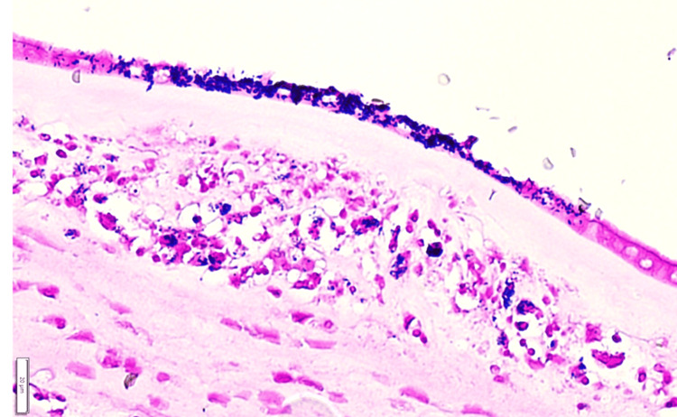
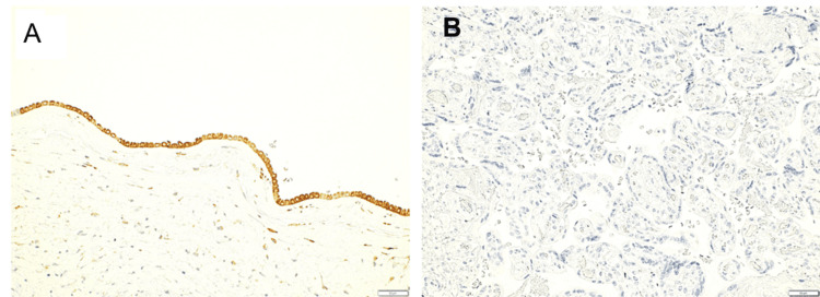
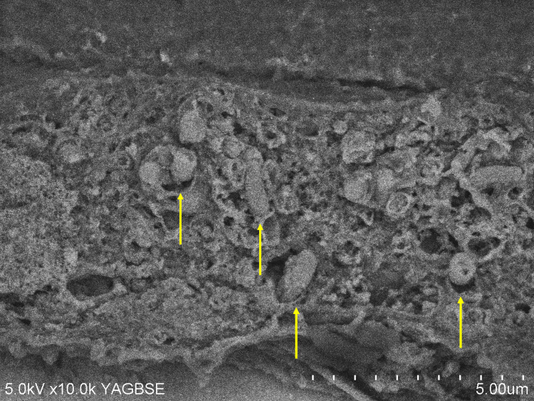
Listeriosis during pregnancy is rare but can lead to premature miscarriage and intrauterine fetal death. A 29-year-old pregnant woman at 29 weeks of gestation was transferred to our hospital because of mild fever and abdominal pain. Emergency cesarean section was performed due to non-reassuring fetal status concomitant with suspected intrauterine infection. An unusually bright yellow amniotic fluid was observed. The oropharyngeal cavity of the neonate was occupied by thick, tenacious yellow mucus, making its removal challenging. The neonate was intubated due to poor oxygenation. Neonatal blood cultures revealed Listeria monocytogenes. Gram staining of cerebrospinal fluid was negative. The neonate was treated and discharged on the 65th day after birth. The mother had a fever of 39.4°C on the first day after surgery; however, no other significant incident occurred. She was discharged on the 10th day after surgery. Placental pathology revealed funisitis, chorioamnionitis, and marginal deciduitis without evidence of villitis, suggesting predominant transvaginal rather than hematogenous infection. Scanning electron microscopy using NanoSuit imaging revealed Listeria monocytogenes in the amniotic epithelium. The presence of Listeria monocytogenes has been reported in foods stored in refrigerators for extended periods of time and in ready-to-eat meals; therefore, it is difficult for pregnant women to be aware of all potential risks. It is important for physicians to recognize that listeriosis may have a long incubation period and present with mild maternal symptoms. Nevertheless, it should be considered as a differential diagnosis. Routine and prompt identification of the causative organism through amniotic fluid and placental swab cultures is important, particularly when intrauterine infection is suspected. Moreover, pathological examination of the placenta can provide insights into the potential route of infection.
Keywords: intrauterine infection; listeria monocytogenes; nanosuit imaging; pregnancy; preterm delivery.
Copyright © 2025, Dohshita et al.
Conflict of interest statement
Human subjects: Informed consent for treatment and open access publication was obtained or waived by all participants in this study. Conflicts of interest: In compliance with the ICMJE uniform disclosure form, all authors declare the following: Payment/services info: All authors have declared that no financial support was received from any organization for the submitted work. Financial relationships: All authors have declared that they have no financial relationships at present or within the previous three years with any organizations that might have an interest in the submitted work. Other relationships: All authors have declared that there are no other relationships or activities that could appear to have influenced the submitted work.
Figures

Similar articles
-
Maternal and neonatal outcomes of elective induction of labor.Evid Rep Technol Assess (Full Rep). 2009 Mar;(176):1-257. Evid Rep Technol Assess (Full Rep). 2009. PMID: 19408970 Free PMC article.
-
Immediate versus deferred delivery of the preterm baby with suspected fetal compromise for improving outcomes.Cochrane Database Syst Rev. 2016 Jul 12;7(7):CD008968. doi: 10.1002/14651858.CD008968.pub3. Cochrane Database Syst Rev. 2016. PMID: 27404120 Free PMC article.
-
Amnioinfusion for chorioamnionitis.Cochrane Database Syst Rev. 2016 Aug 24;2016(8):CD011622. doi: 10.1002/14651858.CD011622.pub2. Cochrane Database Syst Rev. 2016. PMID: 27556818 Free PMC article.
-
[Volume and health outcomes: evidence from systematic reviews and from evaluation of Italian hospital data].Epidemiol Prev. 2013 Mar-Jun;37(2-3 Suppl 2):1-100. Epidemiol Prev. 2013. PMID: 23851286 Italian.
-
Sexual Harassment and Prevention Training.2024 Mar 29. In: StatPearls [Internet]. Treasure Island (FL): StatPearls Publishing; 2025 Jan–. 2024 Mar 29. In: StatPearls [Internet]. Treasure Island (FL): StatPearls Publishing; 2025 Jan–. PMID: 36508513 Free Books & Documents.
References
-
- Detection of Listeria monocytogenes in humans, animals and foods. Iida T, Kanzaki M, Nakama A, Kokubo Y, Maruyama T, Kaneuchi C. J Vet Med Sci. 1998;60:1341–1343. - PubMed
-
- Overview of Listeria monocytogenes contamination in Japan. Okutani A, Okada Y, Yamamoto S, Igimi S. Int J Food Microbiol. 2004;93:131–140. - PubMed