. 2025 Jul 12;17(7):e87802.
doi: 10.7759/cureus.87802. eCollection 2025 Jul.

An Innovative Technique of Revision Surgery for Distal Junctional Failure

Affiliations

An Innovative Technique of Revision Surgery for Distal Junctional Failure

Masato Tanaka et al. Cureus. .

Abstract

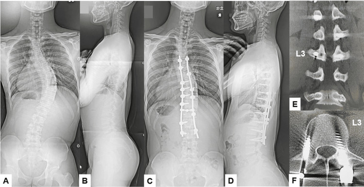
Distal junctional kyphosis (DJK) or the adding-on phenomenon is one of the most challenging complications following long fusion for adolescent idiopathic scoliosis (AIS) or adult spinal deformity (ASD). Among these complications, distal junctional failure (DJF) is defined as a condition requiring revision surgery due to severe symptoms such as intense low back pain, myelopathy, and difficulty in standing and walking. Three patients developed severe lower back pain due to DJF, and two patients underwent revision surgery with a new technique. Surgical outcomes, surgical time, intraoperative blood loss, and operative complications were evaluated. Two patients underwent revision surgery with a novel method for revising distal screw loosening without excessive distal extension. There was no complication, and two patients had solid bony fusion at a two-year follow-up. The revision surgery for type 1 DJK can be challenging because the conventional pedicle screw technique is not feasible for the lower instrumented vertebra, and a more extended distal fusion is required. The adoption of O-arm-guided transdiscal screw fixation has significant clinical implications. This technique also increases accuracy in screw placement, mitigating the risks associated with traditional revision methods and preserving motion segments.

Keywords: c-arm free; distal junctional failure; navigation; reoperation; transdiscal screw.


Conflict of interest statement

Human subjects: Informed consent for treatment and open access publication was obtained or waived by all participants in this study. Ethics Committe of Okayama Rosai Hospital issued approval 543. Animal subjects: All authors have confirmed that this study did not involve animal subjects or tissue. Conflicts of interest: In compliance with the ICMJE uniform disclosure form, all authors declare the following: Payment/services info: All authors have declared that no financial support was received from any organization for the submitted work. Financial relationships: All authors have declared that they have no financial relationships at present or within the previous three years with any organizations that might have an interest in the submitted work. Other relationships: All authors have declared that there are no other relationships or activities that could appear to have influenced the submitted work.

Figures

Figure 1
Figure 1. Patient positioning
Figure 2
Figure 2. Operating room setup
A: O-arm and B: Neuromonitoring.
Figure 3
Figure 3. Muscle dissection
A: Blue arrows indicate the external oblique abdominal muscle, B: Green arrows show the internal oblique abdominal muscle, and C: Red arrows indicate the transverse abdominal muscle. * indicates epidural fat tissue.
Figure 4
Figure 4. Trial and case insertion
A: Coronal image of a trial, B: Axial image of a trial, C: Sagittal image of a trial, D: Coronal image of a cage, E: Axial image of a cage, and E: Sagittal image of a cage. The disc space was carefully expanded using a navigated spreader, and the navigated cage was inserted under navigation guidance.
Figure 5
Figure 5. Pedicle screw insertion
A: Intraoperative image, B: Axial image, and C: Sagittal image. Transdiscal screws should be aimed toward the upper endplate to penetrate the superior endplate.
Figure 6
Figure 6. Schema of the new revision technique for DJK
A: No screw loosening, B: Screw loosening, and C: Revision surgery using transdiscal screws. These transdiscal screws were across the previous screw hole and enhanced the screw pullout strength. DJK: Distal junctional kyphosis
Figure 7
Figure 7. Preoperative images
A: Anteroposterior lumbar radiogram, B: Lateral lumbar radiogram, C: T2-weighted mid sagittal lumbar MRI, D: T2-weighted coronal lumbar MRI, E: T2-weighted axial MRI at T12, and F: T2-weighted axial MRI at T12/L1. Red arrows indicate a large syringomyelia.
Figure 8
Figure 8. Postoperative images
A: Posteroanterior whole spinel radiogram, B: Lateral whole spine radiogram, C: Midsagittal lumbar reconstruction CT, D: Coronal lumbar reconstruction CT, E: Axial CT at L2, and F: Axial CT at L3. Red arrows indicate screw loosening.
Figure 9
Figure 9. Post-revision radiogram
A: Anteroposterior lumbar radiogram and B: Lateral lumbar radiogram.
Figure 10
Figure 10. Final follow-up images
A: Anteroposterior lumbar radiogram, B: Lateral lumbar radiogram, C: Left parasagittal lumbar reconstruction CT, and D: Right parasagittal lumbar reconstruction CT. Red arrows indicate OLIF cages. Block arrows showed transdiscal screws. OLIF: Oblique lateral interbody fusion
Figure 11
Figure 11. Preoperative radiograms
A: Posteroanterior whole spine radiogram, B: Lateral whole spine radiogram, C: Mid sagittal reconstruction CT, and D: 3D CT. Red arrows indicate osteoporotic vertebral fractures.
Figure 12
Figure 12. Postoperative images
A: Anteroposterior lumbar radiogram and B: Lateral lumbar radiogram.
Figure 13
Figure 13. Follow-up images
A: Posteroanterior whole spine radiogram, B: Lateral whole spine radiogram, and C: Mid-sagittal reconstruction CT. The red arrow indicates DJK due to a vertebral fracture. DJK: Distal junctional kyphosis
Figure 14
Figure 14. Pre-and postoperative images
A: Preoperative posteroanterior whole spine radiogram, B: Preoperative lateral whole spine radiogram, C: Postoperative posteroanterior whole spine radiogram, D: Postoperative lateral whole spine radiogram, E: Coronal reconstruction lumbar CT, and F: Axial lumbar CT at L3.  The Cobb angle was corrected from preoperative 56 degrees to postoperative 23 degrees.
Figure 15
Figure 15. Five-year follow-up images
A: Coronal lumbar reconstruction CT, B: Right parasagittal lumbar reconstruction CT, C: Left parasagittal lumbar reconstruction CT, and D and E: Axial CT at L3. Red arrows indicate screw loosening.
Figure 16
Figure 16. Postoperative images after revision surgery
A: Postoperative posteroanterior whole spine radiogram, B: Postoperative lateral whole spine radiogram, C: Right parasagittal lumbar reconstruction CT, D: Left parasagittal lumbar reconstruction CT, and E and F: Axial CT at L3. Red arrows indicate transdiscal screws.

Similar articles

References

    1. Distal junctional kyphosis of adolescent idiopathic thoracic curves following anterior or posterior instrumented fusion: incidence, risk factors, and prevention. Lowe TG, Lenke L, Betz R, et al. Spine (Phila Pa 1976) 2006;31:299–302. - PubMed
    1. Risk factors for distal junctional failure in long-construct instrumentation for adult spinal deformity. McDonnell JM, Evans SR, Ahern DP, et al. Eur Spine J. 2022;31:3654–3661. - PubMed
    1. Risk of distal junctional kyphosis in Scheuermann's kyphosis is decreased by selecting the LIV as two vertebrae distal to the first lordotic disc. Luzzi A, Sardar Z, Cerpa M, et al. Spine Deform. 2022;10:1437–1442. - PubMed
    1. Selective thoracic fusion with segmental pedicle screw fixation in the treatment of thoracic idiopathic scoliosis: more than 5-year follow-up. Suk SI, Lee SM, Chung ER, Kim JH, Kim SS. Spine (Phila Pa 1976) 2005;30:1602–1609. - PubMed
    1. Revision surgery in distal junctional kyphosis. Berjano P, Damilano M, Pejrona M, Langella F, Lamartina C. Eur Spine J. 2020;29:86–102. - PubMed

LinkOut - more resources