. 2025 Jul 28:34:102153.
doi: 10.1016/j.mtbio.2025.102153. eCollection 2025 Oct.

A polymeric nanovesicle delivers sulfopin and gemcitabine to remodel tumor microenvironment for enhanced chemoimmunotherapy against orthotopic pancreatic cancer

Affiliations

A polymeric nanovesicle delivers sulfopin and gemcitabine to remodel tumor microenvironment for enhanced chemoimmunotherapy against orthotopic pancreatic cancer

Weiting Jiang et al. Mater Today Bio. .

Abstract

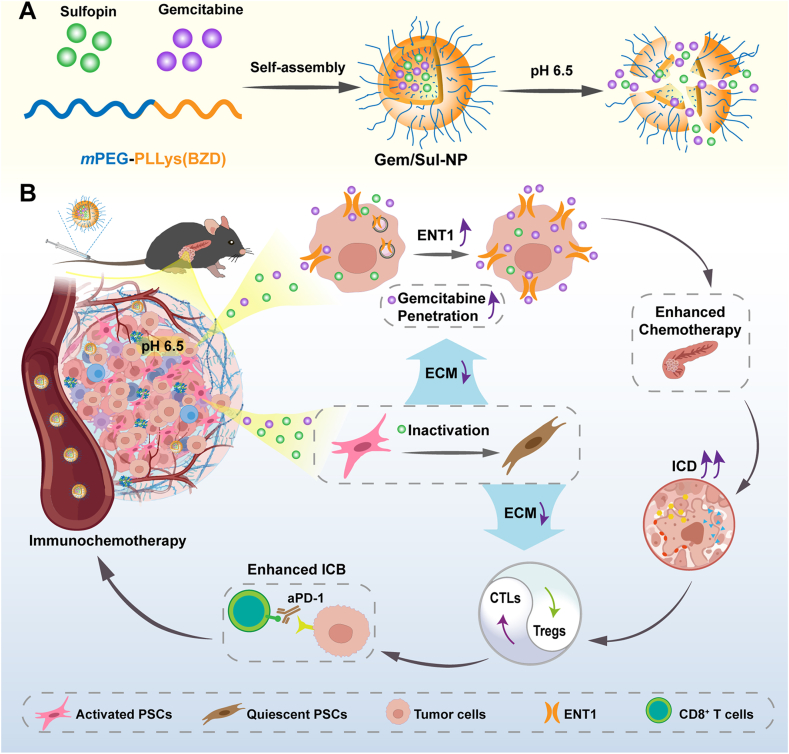
Pancreatic cancers are marked by a highly fibrotic extracellular matrix (ECM) that fosters an immunosuppressive tumor microenvironment (TME), severely limiting the effectiveness of traditional therapies. Emerging evidence suggests that ECM modulation represents a promising strategy to enhance treatment outcomes in pancreatic cancer. Herein, we developed a polymeric nanovesicle for the co-encapsulation and delivery of gemcitabine and prolyl isomerase Pin1 inhibitor sulfopin (Gem/Sul-NP). The engineered Gem/Sul-NP features a compact size of approximately 50 nm, facilitating improved accumulation and penetration within pancreatic tumor tissues. Additionally, the Gem/Sul-NP demonstrates pH-responsive characteristics, undergoing structural disintegration at the acidic pH of 6.5 to achieve controlled drug release in the TME, thereby facilitating synergistic antitumor effects. Sulfopin functions to inhibit pancreatic stellate cells (PSCs) activation, reducing ECM component secretion and disrupting stromal barriers. Besides, it upregulates the expression of the gemcitabine transporter equilibrative nucleoside transporter 1 (ENT1) on tumor cell membranes, thereby enhancing the cellular uptake of gemcitabine and its chemotherapeutic impact. Our in vivo studies confirmed that Gem/Sul-NP treatment effectively remodeled the immunosuppressive TME. When combined with anti-PD-1 therapy, this approach significantly increased CD8+IFNγ+ T cell infiltration, indicating its potential to create favorable conditions for synergistic chemoimmunotherapy against pancreatic cancers.

Keywords: Chemoimmunotherapy; Orthotopic pancreatic cancer; Pancreatic stellate cells; Polymeric nanovesicle; Tumor microenvironment remodeling.


Conflict of interest statement

The authors declare that they have no known competing financial interests or personal relationships that could have appeared to influence the work reported in this paper.

Figures

Image 1
Graphical abstract
Scheme 1
Scheme 1
(A) Schematic illustration of the design and assemble of the nanodrug Gem/Sul-NP. (B) The anti-cancer mechanism of enhanced immunochemotherapy of Gem/Sul-NP.
Fig. 1
Fig. 1
Characterization of the polymer and nanodrug. (A) 1H NMR of mPEG-PLLys(BZD) in DMSO-d6. (B) FTIR spectra of mPEG-PLLys(BZD) and its prepolymers. (C) The particle sizes of the Blank-NP and Gem/Sul-NP. Data are shown as the mean ± SD (n = 3). (D) Transmission electron microscopy (TEM) images of Gem/Sul-NP at pH 7.4 and pH 6.5. (E) The particle size changes of Gem/Sul-NP under various conditions against time. Data are shown as the mean ± SD (n = 3). Samples are stored at 4 °C during non-testing period. (F) In vitro release profiles of gemcitabine from Gem/Sul-NP at pH 7.4 and pH 6.5. Data are shown as the mean ± SD (n = 3).
Fig. 2
Fig. 2
PSC inactivation and chemotherapy potentiation via Pin1 suppression. (A) The western blot result showing the expression of Pin1, α-SMA and collagen I protein level in PSCs after receiving the treatment of Sul-NP at various sulfopin concentrations. (B) CLSM images of immunofluorescence staining for α-SMA in PSCs after treatment with Sul-NP for 72 h. Scale bar: 25 μm. (C) BODIPY 493/503 staining (lipid droplets) in PSCs after treatment with Sul-NP for 72 h. Scale bar: 20 μm. (D) Pin1 and ENT1 protein expression level in Panc02 cells after treatment with Sul-NP for 72 h detected by western-blot. (E) CLSM images of immunofluorescence staining of ENT1 in Panc02 after treatment with Sul-NP for 72 h. Scale bar: 10 μm (Concentration of sulfopin: 20 μM). (F) Relative cell viability of Panc02 cells after incubated with different concentrations of Gem-NP or with Gem/Sul-NP containing 20 μM sulfopin for 72 h. Data are presented as mean ± SD (n = 3). (G) Flow cytometry analysis of Panc02 cell apoptosis after different treatments for 72 h. (H) Statistical analysis of the proportion of apoptosis in (G), the concentration of Sulfopin: 20 μM, Data are presented as mean ± SD. All nanodrugs are pretreated with pH 6.5 for 6 h before use.
Fig. 3
Fig. 3
Biological distribution of nanodrug. (A) Schematic illustrating of nanodrug treatments in vivo. (B) In vivo near-infrared fluorescence tracking of orthotopic pancreatic tumor-bearing mice following intravenous administration of DiR-encapsulated nanovesicles, captured at designated time intervals (DiR label: λex/em = 720/790 nm). (C) Temporal quantification of DiR signal accumulation within tumor regions during the observation period. (D) Postmortem fluorescence evaluation of dissected neoplastic tissues and principal visceral organs 36 h post nanovesicle delivery. (E) Comparative biodistribution analysis of DiR fluorescence signals in the isolated orthotopic tumors and major organs. Data are shown as the mean ± SD (n = 3).
Fig. 4
Fig. 4
Combined antitumor effects of Gem/Sul-NP and aPD-1 in vivo. (A) Schematic illustrating of nanodrug treatments in vivo. Drug dosages: gemcitabine 2.5 mg/kg, sulfopin 4.0 mg/kg, aPD-1 1.2 mg/kg if applied. (B) Magnetic resonance imaging (MRI) showing the tumor size after receiving various treatments including aPD-1, Gem-NP, Sul-NP, Gem/Sul-NP, and Gem/Sul-NP plus aPD-1. Dashed red lines show tumor positions. (C) Body weight change of the mice drug treatment (n = 3). (D) Treatment cohort survival probability analysis in murine pancreatic cancer models (n = 5). (E) TUNEL staining of tumor tissues after treatment. Scale bar: 20 μm. (F) Quantitative Analysis of TUNEL staining. Data are shown as the mean ± SD (n = 3). (For interpretation of the references to colour in this figure legend, the reader is referred to the Web version of this article.)
Fig. 5
Fig. 5
Attenuation of tissue fibrosis. (A) The immunofluorescence staining of α-SMA and Collagen I in tumor tissues. Scale bars: 20 μm. (B) Masson staining of tumor tissues after treatment. Scale bar: 100 μm. (C) Quantitative Analysis of α-SMA and Collagen I after treatment. (D) Quantitative Analysis of Masson after treatment. Data are shown as the mean ± SD (n = 3).
Fig. 6
Fig. 6
The improved tumor microenvironment and enhanced anti-cancer immune responses by nanodrugs. (A) Flow cytometry analysis of CD8+IFNγ+ T cells (gated on CD3+ T cells), CD8+ T cells and CD25+Foxp3+ Tregs (gated on CD3+CD4+ T cells) in tumors after different nanodrug treatments. (B) Quantitative analysis of CD8+IFNγ+ T cells, CD8+ T cells and CD25+Foxp3+ Tregs in tumor tissues after different treatments. Data are shown as the mean ± SD (n = 3). (C) Flow cytometry analysis showing the proportion of intratumoral M1 macrophage (gated on CD11b+F4/80+CD80+) and M2 macrophage (gated on CD11b+F4/80+CD206+) after treatment.

Similar articles

References

    1. Ilic M., Ilic I. Epidemiology of pancreatic cancer. World J. Gastroenterol. 2016;22:9694–9705. doi: 10.3748/wjg.v22.i44.9694. - DOI - PMC - PubMed
    1. Siegel R.L., Miller K.D., Wagle N.S., Jemal A. Cancer statistics. CA Cancer J. Clin. 2023;73:17–48. doi: 10.3322/caac.21763. - DOI - PubMed
    1. Pei Q., Pan J., Zhu H., Ding X., Liu W., Lv Y., Zou X., Luo H. Gemcitabine-treated pancreatic cancer cell medium induces the specific CTL antitumor activity by stimulating the maturation of dendritic cells. Int. Immunopharmacol. 2014;19:10–16. doi: 10.1016/j.intimp.2013.12.022. - DOI - PubMed
    1. Kamisawa T., Wood L.D., Itoi T., Takaori K. Pancreatic cancer. Lancet. 2016;388:73–85. doi: 10.1016/S0140-6736(16)00141-0. - DOI - PubMed
    1. Shui L., Cheng K., Li X., Shui P., Zhou X., Li J., Yi C., Cao D. Study protocol for an open-label, single-arm, phase Ib/II study of combination of toripalimab, nab-paclitaxel, and gemcitabine as the first-line treatment for patients with unresectable pancreatic ductal adenocarcinoma. BMC Cancer. 2020;20:636. doi: 10.1186/s12885-020-07126-3. - DOI - PMC - PubMed

LinkOut - more resources