. 2025 Jul 29:34:102150.
doi: 10.1016/j.mtbio.2025.102150. eCollection 2025 Oct.

Sustained release of cathepsin D/cathepsin K from injectable hydrogel microspheres in anti-photoaging

Affiliations

Sustained release of cathepsin D/cathepsin K from injectable hydrogel microspheres in anti-photoaging

Wenyi Hou et al. Mater Today Bio. .

Abstract

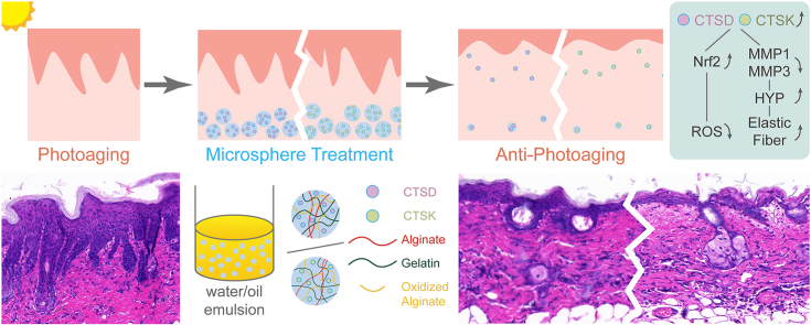
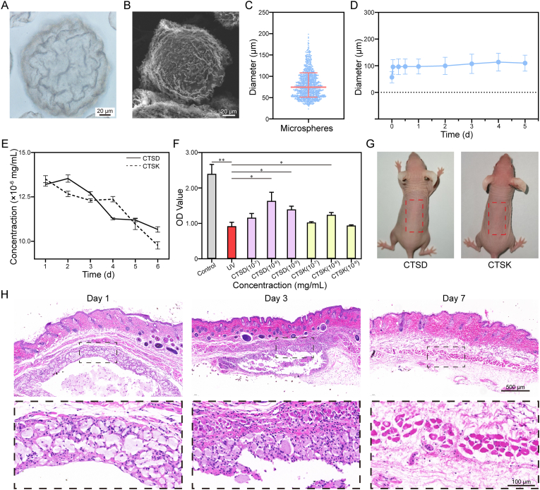
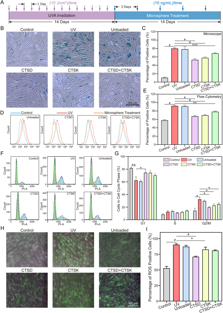
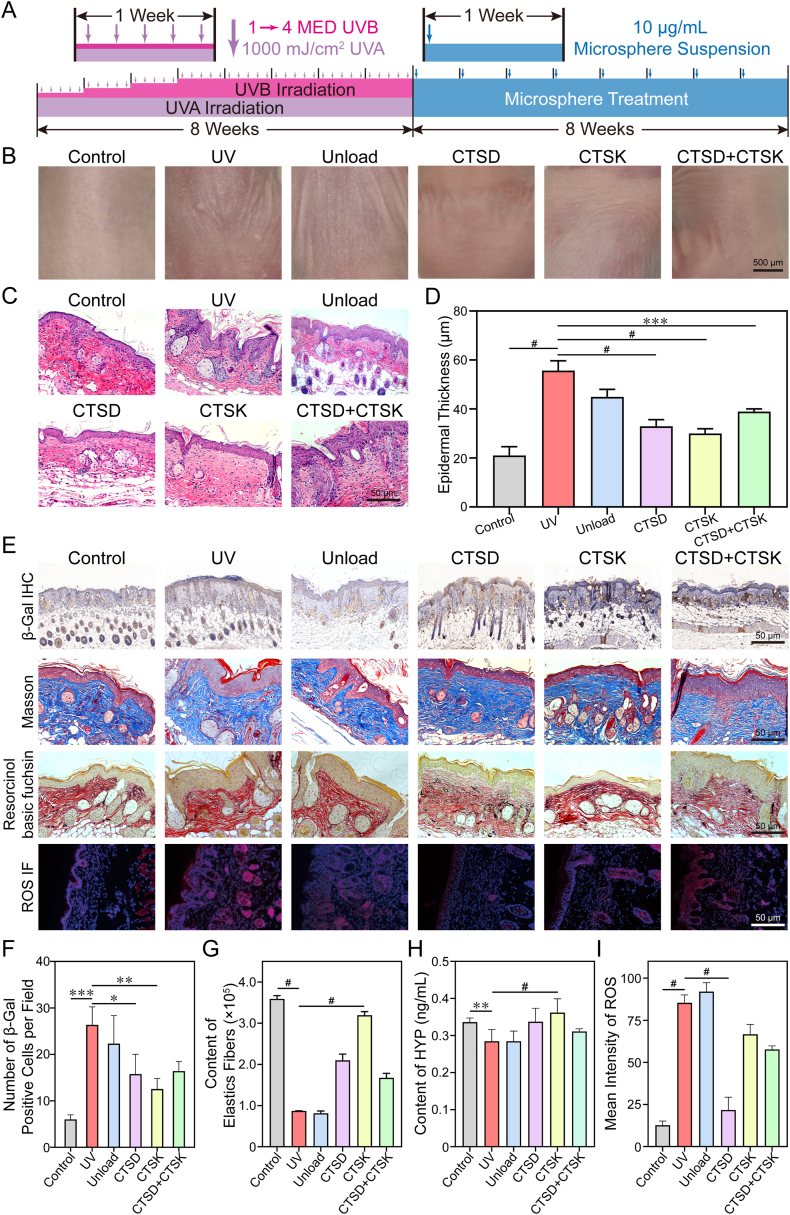
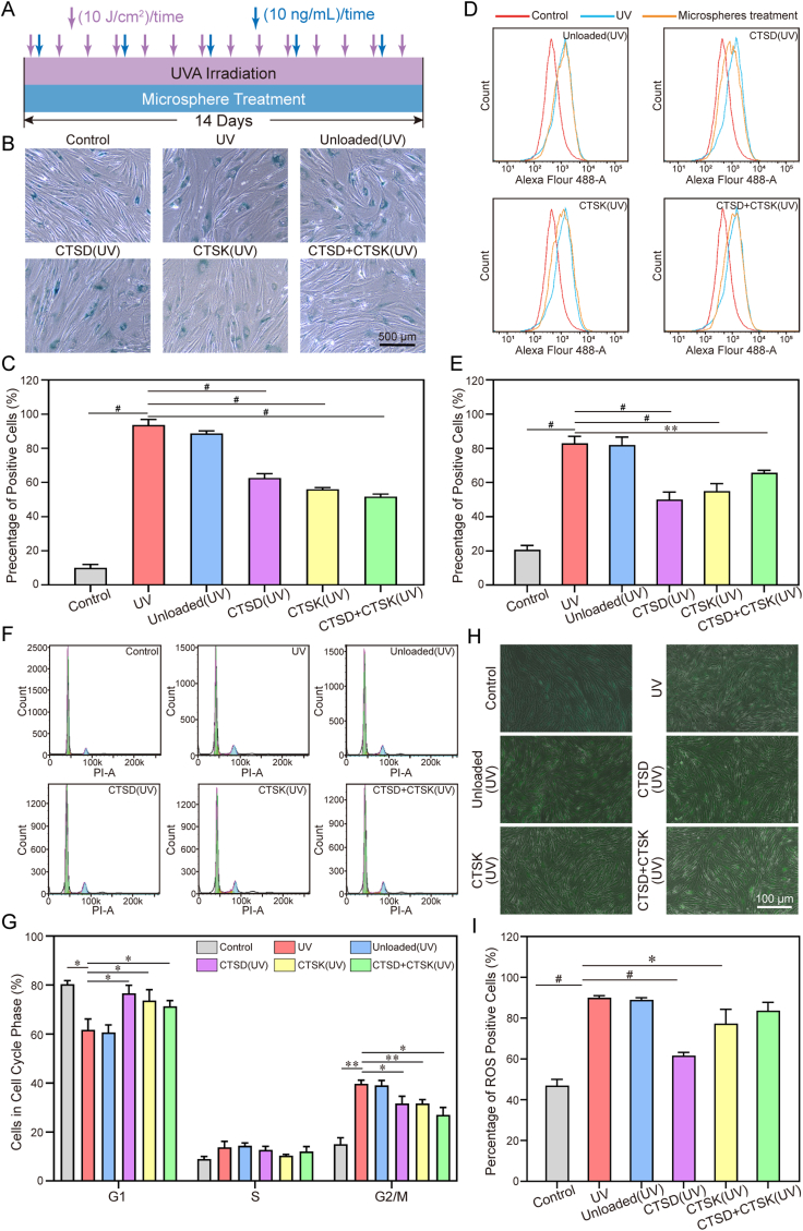
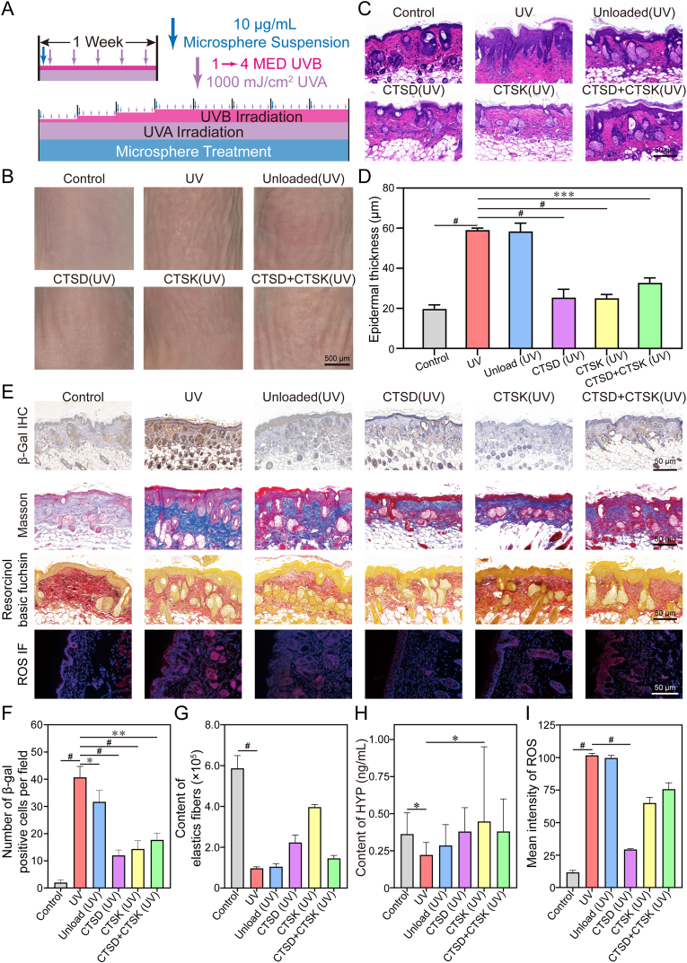
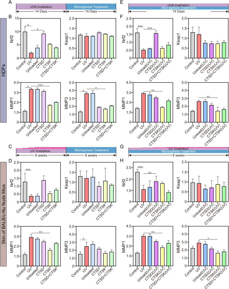
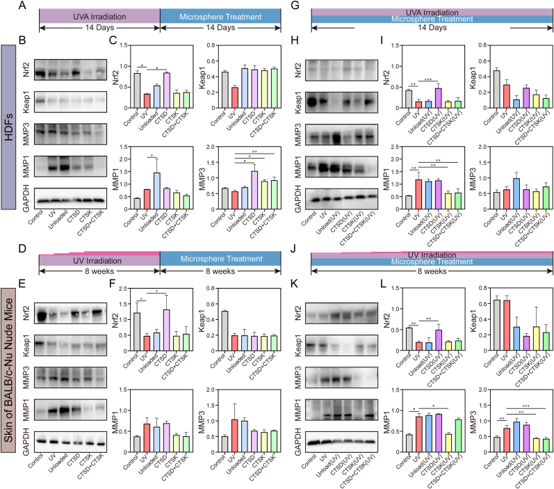
There have been various interventions for photoaging skin, such as retinoic acid treatment and laser therapy. However, more precise, secure, and effective technologies and materials were still need to be explored. Cathepsin D (CTSD) repairs the epidermal barrier in chronic photodamaged skin through increasing TGase-1 expression and activity. Cathepsin K (CTSK) affects the metabolism of skin elastic fibers. Therefore, gelatin/alginate composite hydrogel microspheres loaded with CTSD or CTSK were fabricated. The microspheres with an average size of 79.8 ± 30.4 μm were stable in PBS (pH 7.4) for 5 days and then disintegrated in vitro. 0.1 mg/mL of microspheres continuously released 12-13 ng/mL CTSD or CTSK for the first three consecutive days. In vivo, the microspheres were almost completed degraded on day 7 after the subcutaneous injection. The biosafety and efficacy of CTSD- and CTSK- microspheres were confirmed in vitro and in vivo. They reduced ROS and inhibited the degradation of skin collagen and elastic fibers by upregulating Nrf2 and downregulating MMP-1 and MMP-3, which reversed photodamage of skin induced by UV. In conclusion, the gelatin/alginate composite microspheres loaded with CTSD or CTSK showed a great potential for chronic photodamaged skin.

Keywords: Cathepsin D; Cathepsin K; Degradable microspheres; Photoaging; Skin.


Conflict of interest statement

The authors declare the following financial interests/personal relationships which may be considered as potential competing interests: Yue Zheng reports financial support was provided by National Natural Science Foundation of China. Yue Zheng reports financial support was provided by Science and Technology Program of Guangzhou. Yue Zheng reports financial support was provided by 10.13039/501100003453Natural Science Foundation of Guangdong Province. Yue Zheng has patent Enzyme-Loaded Controlled-Release Microspheres for Skin Photoaging Prevention/Treatment: Preparation Method and Applications pending to Yue Zheng. If there are other authors, they declare that they have no known competing financial interests or personal relationships that could have appeared to influence the work reported in this paper.

Figures

Image 1
Graphical abstract
Fig. 1
Fig. 1
Characterization of microspheres. A. Light microscopy image and B. scanning electron microscope (SEM) of a microsphere. C. Microsphere diameter distribution. D. Change of average diameter for microspheres with time after rehydration. E. Release curve of enzyme-loaded microspheres. F. The appropriate concentration of CTSD and CTSK for in vitro studies was selected by cck8 assay (10−8 mg/mL) (n = 3; data = mean ± SD; ∗P < 0.05, ∗∗P < 0.01 compared to UV group). G. The back skin of nude mice after subcutaneous injection of microspheres at 10 μg/mL. H.In vivo degradation of microspheres; 7 days after injection, the microspheres were degraded completely.
Fig. 2
Fig. 2
Co-culture of HDFs with microspheres. A. Timeline of the treatment. Chronic photodamaged cell model was established via UV radiation for 14 d and then cells were treated with microspheres without further UV exposure. B. Morphology and SA-β-gal staining shown by optical microscopy. C. Percentage of SA-β-gal positive cells in the corresponding light microscope images. Flow cytometry results for D. E. cell senescence, F. G. cell cycle. H. I. Immunofluorescence staining showing ROS content in fibroblasts after microsphere treatment. (n = 3; data = mean ± SD; ∗P < 0.05, ∗∗P < 0.01, ∗∗∗P < 0.001, #P < 0.0001 indicated a significant difference.)
Fig. 3
Fig. 3
Subcutaneous injection of microspheres to evaluate the effect against photoaging. A. Timeline of the treatment. Chronic photodamaged animal model was created via UV exposure for 8 weeks; microspheres were injected every week which lasted for 8 weeks without UV exposure. B. Digital images of the dorsal skins. C. H&E staining images of photodamaged skins. D. Epidermal thickness calculated from the H&E image. E. Histological assessment of skin chronic photodamage and β-gal expression using Masson staining, resorcinol basic fuchsin staining and immunofluorescence staining of ROS. F. Number of β-gal positive cells per field. G. Content of elastic fibers. H. Content of HYP. I. Mean intensity of ROS. (n = 3; data = mean ± SD; ∗P < 0.05, ∗∗P < 0.01, ∗∗∗P < 0.001, #P < 0.0001 indicated a significant difference).
Fig. 4
Fig. 4
In vitro co-culture of HDFs with microsphere to mimic daily UV exposure and microsphere treatment. A. Treatment timeline. B. Light microscope image for SA-β-gal staining and C. the percentage of corresponding SA-β-gal positive cells. Flow cytometry results for D. E. cell senescence and F. G. cell cycle. H, I. Immunofluorescence staining showing ROS content in fibroblasts. (n = 3; data = mean ± SD; ∗P < 0.05, ∗∗P < 0.01, ∗∗∗P < 0.001, #P < 0.0001 indicated a significant difference).
Fig. 5
Fig. 5
In vivo evaluation of microspheres against photoaging. To mimic daily life, UV exposure and subcutaneous injection of microspheres were performed at the same time. A. Treatment timeline. B. Dorsal skin of mice. C. H&E staining and D. corresponding epidermal thickness. E. Histological assessment of skin chronic photodamage and β-gal expression. F. Number of β-gal positive cells per field. G. Content of elastic fibers. H. Content of HYP. I. Mean intensity of ROS. (n = 3; data = mean ± SD; ∗P < 0.05, ∗∗P < 0.01, ∗∗∗P < 0.001, #P < 0.0001 indicated a significant difference).
Fig. 6
Fig. 6
qPCR results of HDFs and mice dorsal skin showed that Nrf2 was upregulated in CTSD group, MMP-1, MMP-3 were downregulated in CTSK group. Treatment timeline for A. E.in vitro and C. G.in vivo tests. B. F. qPCR results of HDFs. D. H. qPCR results of mice dorsal skin. (n = 3; data = mean ± SD; ∗P < 0.05, ∗∗P < 0.01, ∗∗∗P < 0.001, #P < 0.0001 indicated a significant difference.
Fig. 7
Fig. 7
Western blot results of HDFs and mice dorsal skin showed that Nrf2 was upregulated in CTSD group, MMP-1, MMP-3 were downregulated in CTSK group. Treatment timeline for A. G.in vitro and D. J.in vivo tests. Western blot results of B. C. H. I. HDFs and E. F. K. L. mice dorsal skin. (n = 3; data = mean ± SD; ∗P < 0.05, ∗∗P < 0.01, ∗∗∗P < 0.001, #P < 0.0001 indicated a significant difference.

Similar articles

References

    1. Gruber F., Kremslehner C., Eckhart L., Tschachler E. Cell aging and cellular senescence in skin aging - recent advances in fibroblast and Keratinocyte biology. Exp. Gerontol. 2020;130 doi: 10.1016/j.exger.2019.110780. - DOI - PubMed
    1. Gromkowska-Kępka K.J., Puścion-Jakubik A., Markiewicz-Żukowska R., Socha K. The impact of ultraviolet radiation on skin photoaging - review of in vitro studies. J. Cosmet. Dermatol. 2021;20(11):3427. doi: 10.1111/jocd.14033. - DOI - PMC - PubMed
    1. Mostafa D.K., Nayel O.A., Abdulmalek S., Abdelbary A.A., Ismail C.A. Modulation of autophagy, apoptosis and oxidative stress: a clue for repurposing metformin in photoaging. Inflammopharmacology. 2022;30(6):2521. doi: 10.1007/s10787-022-01041-8. - DOI - PMC - PubMed
    1. Salminen A., Kaarniranta K., Kauppinen A. Photoaging: UV radiation-induced inflammation and immunosuppression accelerate the aging process in the skin. Inflamm. Res. 2022;71(7–8):817. doi: 10.1007/s00011-022-01598-8. - DOI - PMC - PubMed
    1. Shen Y.J., Kim K., Zhu Z.X., Zhang S.Y., Jiang M.Y., Liu Z.L., Zheng Y.Y., Li X.K., Jin L.T., Cong W.T. δ-Catenin requirement in keratinocyte proliferation and DNA repair identifies a therapeutic target for photoaging. J. Invest. Dermatol. 2023;143(1):26. doi: 10.1016/j.jid.2022.07.009. - DOI - PubMed

LinkOut - more resources