. 2024 Nov 12:2:imag-2-00360.
doi: 10.1162/imag_a_00360. eCollection 2024.

Functional localization of the human auditory and visual thalamus using a thalamic localizer functional magnetic resonance imaging task

Affiliations

Functional localization of the human auditory and visual thalamus using a thalamic localizer functional magnetic resonance imaging task

John C Williams et al. Imaging Neurosci (Camb). .

Abstract

Functional magnetic resonance imaging (fMRI) of the auditory and visual sensory systems of the human brain is an active area of investigation in the study of human health and disease. The medial geniculate nucleus (MGN) and lateral geniculate nucleus (LGN) are key thalamic nuclei involved in the processing and relay of auditory and visual information, respectively, and are the subject of blood-oxygen-level-dependent (BOLD) fMRI studies of neural activation and functional connectivity in human participants. However, localization of BOLD fMRI signal originating from neural activity in MGN and LGN remains a technical challenge, due, in part, to the poor definition of boundaries of these thalamic nuclei in standard T1-weighted and T2-weighted magnetic resonance imaging sequences. Here, we report the development and evaluation of an auditory and visual sensory thalamic localizer (TL) fMRI task that produces participant-specific functionally-defined regions of interest (fROIs) of both MGN and LGN, using 3 Tesla multiband fMRI and a clustered-sparse temporal acquisition sequence, in less than 16 minutes of scan time. We demonstrate the use of MGN and LGN fROIs obtained from the TL fMRI task in standard resting-state functional connectivity (RSFC) fMRI analyses in the same participants. In RSFC analyses, we validated the specificity of MGN and LGN fROIs for signals obtained from primary auditory and visual cortex, respectively, and benchmarked their performance against alternative atlas- and segmentation-based localization methods. The TL fMRI task and analysis code (written in Presentation and MATLAB, respectively) have been made freely available to the wider research community.

Keywords: auditory processing; functional localizer task; lateral geniculate; medial geniculate; resting-state functional connectivity; visual processing.


Conflict of interest statement

A.A.-D. received consulting fees and/or honoraria from Sunovion Pharmaceuticals Inc., Otsuka Pharmaceutical Co., Ltd., Merck & Co., Inc., Neurocrine Biosciences, Inc., F. Hoffmann-La Roche AG, and C.H. Boehringer Sohn AG & Co. KG. A.A.-D. holds stock options in Herophilus, Inc. and in Terran Biosciences, Inc. All other authors declare that they have no known competing financial interests or personal relationships that could have influenced or appear to have influenced the work reported in this paper.

Figures

Fig. 1.
Fig. 1.
The sensory thalamic localizer (TL) task. A schematic diagram of a TL task run is shown in panel (A). Each run begins with a single 12-second fixation trial (green), followed by sixteen 12-second stimulus trials, consisting of 8 auditory and 8 visual trials (blue), presented in pseudorandom order, using clustered-sparse temporal acquisition. Sample auditory and visual trials are respectively shown in panels (B and C). Each trial consists of 9 seconds of stimulation, followed by a single acquisition cluster of 3 volumes (orange). Acquisition clusters are 3 TRs in duration, totaling 2.55 seconds in NYSPI data and 2.4 seconds in SBU data. Stimuli are immediately preceded and succeeded by gaps of equal duration, during which no stimulus is presented, in order to bring the total trial time to 12 seconds; this gap time is 225 ms in NYSPI data and 300 ms in SBU data. Auditory trials consist of nine sets of 900 ms amplitude-normalized snippets of instrumental music presented in pseudorandom order and separated by 100 ms gaps. Visual trials consist of a circular checkerboard alternating between black and white at 7.5 Hz. Participants were asked to complete a total of four task runs.
Fig. 2.
Fig. 2.
Group-level probability density maps of medial geniculate nucleus (MGN) and lateral geniculate nucleus (LGN) functionally-defined regions of interest (fROIs) obtained from the sensory thalamic localizer (TL) task (A, C, E, G) and violin plots of ROI size (B, D, F, H) for left MGN (A, B), right MGN (C, D), left LGN (E, F), and right LGN (G, H). Crosshair coordinates for each view are displayed with each set of images.
Fig. 3.
Fig. 3.
Thresholded (binarized) group-level probability density maps of (A, B) left and right medial geniculate nucleus (MGN, blue) and (C, D) lateral geniculate nucleus (LGN, yellow) functionally-defined regions of interest (fROIs) obtained from the sensory thalamic localizer (TL) task. Probability density maps for MGN and LGN were binarized by thresholding at 50% of participants. Image views and crosshairs are centered on the centroid of each fROI in Montreal Neurological Institute (MNI) 152 non-linear 6th-generation (MNI152NLin6), with crosshair coordinates shown for each set of images.
Fig. 4.
Fig. 4.
Evaluation of medial geniculate nucleus (MGN) and lateral geniculate nucleus (LGN) functionally-defined regions of interest (fROIs) obtained from the sensory thalamic localizer (TL) task. Pairwise resting-state function connectivity (RSFC) was evaluated between TL-derived geniculi and standard auditory cortex (AC) and visual cortex (VC) ROIs derived from the FreeSurfer atlas (f). (A) Connectivity between MGN and AC (MGN-AC), MGN and VC (MGN-VC), LGN and AC (LGN-AC), and LGN and VC (LGN-VC). (B) Selectivity of MGN and LGN ROIs for AC and VC, respectively, measured as within-participant differences in the RSFC values shown in panel A. MGN-AC minus MGN-VC quantifies MGN selectivity for AC over VC; MGN-AC minus LGN-AC quantifies AC selectivity for MGN over LGN. Likewise, LGN-VC minus MGN-VC quantifies VC selectivity for MGN over LGN, and LGN-VC minus LGN-AC quantifies LGN selectivity for VC over AC. p-values for significance of sample-wide mean difference are shown; asterisks (*) denote significance after false discovery rate correction (α = 0.05, one-tailed, one-sample t-test).
Fig. 5.
Fig. 5.
Benchmarking of medial geniculate nucleus (MGN) and lateral geniculate nucleus (LGN) functionally-defined regions of interest (fROIs) obtained from the sensory thalamic localizer (TL) task through resting-state functional connectivity (RSFC) with auditory cortex (AC) and visual cortex (VC) ROIs, respectively. AC and VC regions of interest (ROIs) were obtained from the FreeSurfer (FS) Desikan-Killiany atlas (Atlas_wmparc.2.nii.gz). MGN and LGN derived from the TL task are evaluated relative to those derived from (1) MNI space atlas ROIs from the Wake Forest University (WFU) PickAtlas, (2) THOMAS segmentation applied to each subject’s T1w image, and (3) the FreeSurfer thalamic segmentation applied to each subject’s T1w. Panel (A) shows MGN-AC RSFC using the TL MGN ROI alongside the three named alternatives; panel (B) shows the within-participant differences between MGN-AC connectivity from the TL-derived MGNs and the three alternatives (i.e., differences between TL and other values in panel A). Likewise, panel (C) shows LGN-VC RSFC using the TL LGN ROI alongside alternatives derived from WFU, THOMAS, and FS; panel (D) shows the within-participant differences between TL connectivity and the three alternatives shown in panel (C). p-values for significance of sample-wide mean difference are shown; asterisks (*) denote significance (α < 0.05, two-tailed, one-sample t-test).
Fig. 6.
Fig. 6.
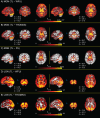
Whole-brain T-statistic maps for differences in seed connectivity between thalamic localizer (TL) derived medial geniculate nucleus (MGN) and lateral geniculate nucleus (LGN) functionally-defined regions of interest (fROIs), relative to seed connectivity calculated from three alternative regions of interest (ROIs): Wake Forest University PickAtlas (WFU), the Thalamus Optimized Multi Atlas Segmentation (THOMAS), and the FreeSurfer thalamic nuclei segmentation (FS). T-statistics are thresholded at α = 0.05 (two-tailed, family-wise error rate corrected). Image views are centered on either auditory cortex (panels A-C) or visual cortex (panels D and E) of each hemisphere, with crosshair coordinates for each view shown. A panel for LGN TL − FS T-statistics is not shown, as there were no significant voxels.

Update of

References

    1. Alberton , B. A. V. , Nichols , T. E. , Gamba , H. R. , & Winkler , A. M. ( 2020. ). Multiple testing correction over contrasts for brain imaging . Neuroimage , 216 , 116760 . 10.1016/j.neuroimage.2020.116760 - DOI - PMC - PubMed
    1. Almasabi , F. , Janssen , M. L. F. , Devos , J. , Moerel , M. , Schwartze , M. , Kotz , S. A. , Jahanshahi , A. , Temel , Y. , & Smit , J. V. ( 2022. ). The role of the medial geniculate body of the thalamus in the pathophysiology of tinnitus and implications for treatment . Brain Res , 1779 , 147797 . 10.1016/j.brainres.2022.147797 - DOI - PubMed
    1. American Psychiatric Association . ( 2000. ). Diagnostic and statistical manual of mental disorders: DSM-IV-TR ( 4th ed., text revision. ed.). American Psychiatric Association; . ISBN: 9780890420621. https://books.google.co.in/books/about/Diagnostic_and_Statistical_Manual...
    1. American Psychiatric Association . ( 2013. ). Diagnostic and statistical manual of mental disorders: DSM-5 ( 5th ed.). American Psychiatric Association; . 10.1176/appi.books.9780890425596 - DOI
    1. Andrews , T. J. , Halpern , S. D. , & Purves , D. ( 1997. ). Correlated size variations in human visual cortex, lateral geniculate nucleus, and optic tract . J Neurosci , 17 ( 8 ), 2859 – 2868 . 10.1523/JNEUROSCI.17-08-02859.1997 - DOI - PMC - PubMed

LinkOut - more resources