

Unilateral Painless Visual Loss in Sphenoid Mucoceles with Optic Neuropathy
- PMID: 40800573
- PMCID: PMC12342703
- DOI: 10.1159/000546758
Unilateral Painless Visual Loss in Sphenoid Mucoceles with Optic Neuropathy
Abstract
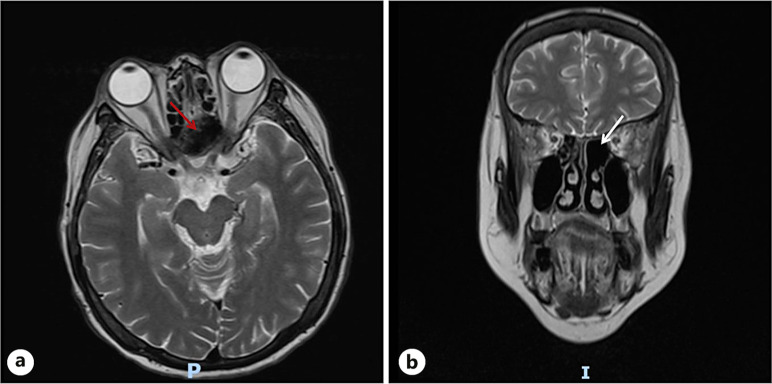
Introduction: Mucoceles are benign, slow-growing cystic formations located within the paranasal sinuses, caused by complete ostial obstruction and accumulation of mucous secretions. Here, we report a case of a patient who initially presented with unilateral painless visual impairment and was ultimately diagnosed with bilateral sphenoid sinus mucoceles (SSMs) after two hospitalizations.
Case presentation: A 67-year-old woman presented with a 7-day history of decreased vision in her left eye. She denied any orbital pain, headache, or restricted eye movement and was diagnosed with retrobulbar ischemic optic neuropathy on the first admission. After drug treatment, the visual acuity of patient improved significantly, but 3 months after discharge, the patient was hospitalized again due to recurrent vision loss accompanied by left orbital pain and left temporal pulsatile headaches. After multiple imaging examinations, the patient was ultimately diagnosed with SSMs and her visual acuity was restored after surgical treatment.
Conclusions: The majority of SSMs are associated with ocular symptoms, with only a minority presenting solely with unilateral or bilateral vision loss, as exemplified in this case. Therefore, understanding the clinical features of visual disturbances secondary to SSMs is crucial to aiding more prompt diagnosis and treatment.
Keywords: Compressive optic neuropathy; Sphenoid sinus mucoceles; Visual impairment.
© 2025 The Author(s). Published by S. Karger AG, Basel.
Conflict of interest statement
The authors have no conflicts of interest to declare.
Figures


References
-
- Makihara S, Kariya S, Okano M, Naito T, Tsumura M, Nishizaki K. Orbital complications of infected mucocele in the paranasal sinuses. Auris Nasus Larynx. 2020;47(6):990–5. - PubMed
-
- Peng X, Yue ZZ, Zhang YL, Sun PY. Nasal sinus mucoceles manifesting ocular symptoms. J Craniofac Surg. 2023;34(2):e141–5. - PubMed
-
- Stankiewicz JA. Sphenoid sinus mucocele. Arch Otolaryngol Head Neck Surg. 1989;115(6):735–40. - PubMed
Publication types
LinkOut - more resources
Full Text Sources
