Surgical management of large facial congenital melanocytic nevi using Ahmad technique: a case report
- PMID: 40800642
- PMCID: PMC12343088
- DOI: 10.1093/jscr/rjaf612
Surgical management of large facial congenital melanocytic nevi using Ahmad technique: a case report
Abstract
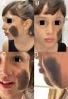
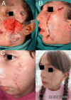
We report the case of a 9-year-old girl with a large congenital hairy pigmented nevus on her left cheek causing psychological distress. A novel and promising surgical method called the 'Ahmad Technique,' named after the author of this manuscript, was employed. This approach uses a single tissue expander in a structured four-stage treatment plan. Initially, a 30 ml subcutaneous expander was inserted via a preauricular incision and gradually inflated every 10 days over 4 months. After complete deflation, a partial excision of the nevus was performed, with histopathology confirming a benign intradermal melanocytic nevus. Following wound healing, tissue expansion resumed for another four months. The third stage included a second partial excision followed by a final expansion phase. In the fourth stage, complete excision of the nevus and expander capsule was done, and reconstruction used the expanded skin flap. The patient achieved excellent aesthetic and psychological outcomes, confirming the Ahmad Technique as a safe, effective, and innovative option for complex facial congenital melanocytic nevi.
Keywords: Ahmad technique; congenital melanocytic nevus; facial reconstruction; tissue expansion.
© The Author(s) 2025. Published by Oxford University Press and JSCR Publishing Ltd.
Conflict of interest statement
None declared.
Figures


References
-
- Tannous ZS, Mihm MC Jr. Congenital melanocytic nevi: clinical and histopathologic features, risk of melanoma, and clinical management. Dermatol Surg 2005;31:1135–40. - PubMed
-
- Arad E, Zuker RM. Giant congenital melanocytic nevi: a plastic surgery perspective. Clin Plast Surg 2011;38:253–67.
Publication types
LinkOut - more resources
Full Text Sources

