Body size and intracranial volume interact with the structure of the central nervous system: A multi-center in vivo neuroimaging study
- PMID: 40800833
- PMCID: PMC12319740
- DOI: 10.1162/imag_a_00559
Body size and intracranial volume interact with the structure of the central nervous system: A multi-center in vivo neuroimaging study
Abstract
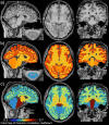
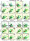
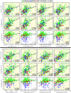
Clinical research emphasizes the implementation of rigorous and reproducible study designs that rely on between-group matching or controlling for sources of biological variation such as subject's sex and age. However, corrections for body size (i.e., height and weight) are mostly lacking in clinical neuroimaging designs. This study investigates the importance of body size parameters in their relationship with spinal cord (SC) and brain magnetic resonance imaging (MRI) metrics. Data were derived from a cosmopolitan population of 267 healthy human adults (age 30.1 ± 6.6 years old, 125 females). We show that body height correlates with brain gray matter (GM) volume, cortical GM volume, total cerebellar volume, brainstem volume, and cross-sectional area (CSA) of cervical SC white matter (CSA-WM; 0.44 ≤ r ≤ 0.62). Intracranial volume (ICV) correlates with body height (r = 0.46) and the brain volumes and CSA-WM (0.37 ≤ r ≤ 0.77). In comparison, age correlates with cortical GM volume, precentral GM volume, and cortical thickness (-0.21 ≥ r ≥ -0.27). Body weight correlates with magnetization transfer ratio in the SC WM, dorsal columns, and lateral corticospinal tracts (-0.20 ≥ r ≥ -0.23). Body weight further correlates with the mean diffusivity derived from diffusion tensor imaging (DTI) in SC WM (r = -0.20) and dorsal columns (-0.21), but only in males. CSA-WM correlates with brain volumes (0.39 ≤ r ≤ 0.64), and with precentral gyrus thickness and DTI-based fractional anisotropy in SC dorsal columns and SC lateral corticospinal tracts (-0.22 ≥ r ≥ -0.25). Linear mixture of age, sex, or sex and age, explained 2 ± 2%, 24 ± 10%, or 26 ± 10%, of data variance in brain volumetry and SC CSA. The amount of explained variance increased to 33 ± 11%, 41 ± 17%, or 46 ± 17%, when body height, ICV, or body height and ICV were added into the mixture model. In females, the explained variances halved suggesting another unidentified biological factor(s) determining females' central nervous system (CNS) morphology. In conclusion, body size and ICV are significant biological variables. Along with sex and age, body size should therefore be included as a mandatory variable in the design of clinical neuroimaging studies examining SC and brain structure; and body size and ICV should be considered as covariates in statistical analyses. Normalization of different brain regions with ICV diminishes their correlations with body size, but simultaneously amplifies ICV-related variance (r = 0.72 ± 0.07) and suppresses volume variance of the different brain regions (r = 0.12 ± 0.19) in the normalized measurements.
Keywords: body height and weight; brain; in vivo human neuroimaging; intracranial volume; spinal cord; structural magnetic resonance imaging.
© 2025 The Authors. Published under a Creative Commons Attribution 4.0 International (CC BY 4.0) license.
Conflict of interest statement
Since June 2022, Dr. A.K. Smith has been employed by GE HealthCare. This article was co-authored by Dr. Smith in his personal capacity. The opinions expressed in the article are his and do not necessarily reflect the views of GE HealthCare. Since August 2022, Dr. M. M. Laganà has been employed by Canon Medical Systems srl, Rome, Italy. This article was co-authored by Dr. M. M. Laganà in her personal capacity. The opinions expressed in the article are her own and do not necessarily reflect the views of Canon Medical Systems. Since September 2023, Dr. Papp has been an employee of Siemens Healthcare AB, Sweden. This article was co-authored by Dr. Papp in his personal capacity. The views and opinions expressed in this article are his own and do not necessarily reflect the views of Siemens Healthcare AB, or Siemens Healthineers AG. Since January 2024, Dr. Barry has been employed by the National Institute of Biomedical Imaging and Bioengineering at the NIH. This article was co-authored by Robert Barry in his personal capacity. The opinions expressed in the article are his own and do not necessarily reflect the views of the NIH, the Department of Health and Human Services, or the United States government. Guillaume Gilbert is an employee of Philips Healthcare. S Llufriu received compensation for consulting services and speaker honoraria from Biogen Idec, Novartis, Bristol Myer Squibb Genzyme, Sanofi Jansen, and Merck. The Max Planck Institute for Human Cognitive and Brain Sciences and Wellcome Centre for Human Neuroimaging have institutional research agreements with Siemens Healthcare. N.W. holds a patent on acquisition of MRI data during spoiler gradients (US 10,401,453 B2). N.W. was a speaker at an event organized by Siemens Healthcare and was reimbursed for the travel expenses. C.G.W.K. is a shareholder in Queen Square Analytics Ltd. M.D. is a shareholder in Imeka Solutions. The other authors declare no competing interests.
Figures


Update of
-
Body size interacts with the structure of the central nervous system: A multi-center in vivo neuroimaging study.bioRxiv [Preprint]. 2024 May 1:2024.04.29.591421. doi: 10.1101/2024.04.29.591421. bioRxiv. 2024. Update in: Imaging Neurosci (Camb). 2025 May 07;3:imag_a_00559. doi: 10.1162/imag_a_00559. PMID: 38746371 Free PMC article. Updated. Preprint.
References
-
- Ahn , J. H. , Kim , M. , Kim , J. S. , Youn , J. , Jang , W. , Oh , E. , Lee , P. H. , Koh , S.-B. , Ahn , T.-B. , & Cho , J. W. ( 2019. ). Midbrain atrophy in patients with presymptomatic progressive supranuclear palsy-Richardson’s syndrome . Parkinsonism & Related Disorders , 66 , 80 – 86 . 10.1016/j.parkreldis.2019.07.009 - DOI - PubMed
-
- Alfaro-Almagro , F. , Jenkinson , M. , Bangerter , N. K. , Andersson , J. L. R. , Griffanti , L. , Douaud , G. , Sotiropoulos , S. N. , Jbabdi , S. , Hernandez-Fernandez , M. , Vallee , E. , Vidaurre , D. , Webster , M. , McCarthy , P. , Rorden , C. , Daducci , A. , Alexander , D. C. , Zhang , H. , Dragonu , I. , Matthews , P. M. , … Smith , S. M. ( 2018. ). Image processing and Quality Control for the first 10,000 brain imaging datasets from UK Biobank . NeuroImage , 166 , 400 – 424 . 10.1016/j.neuroimage.2017.10.034 - DOI - PMC - PubMed
Grants and funding
- P41 EB027061/EB/NIBIB NIH HHS/United States
- R00 EB016689/EB/NIBIB NIH HHS/United States
- K23 NS104211/NS/NINDS NIH HHS/United States
- P30 NS076408/NS/NINDS NIH HHS/United States
- R01 NS133305/NS/NINDS NIH HHS/United States
- R61 NS118651/NS/NINDS NIH HHS/United States
- R01 NS128478/NS/NINDS NIH HHS/United States
- R21 EB031211/EB/NIBIB NIH HHS/United States
- R01 NS109450/NS/NINDS NIH HHS/United States
- K24 NS126781/NS/NINDS NIH HHS/United States
- K01 EB030039/EB/NIBIB NIH HHS/United States
- R03 NS139000/NS/NINDS NIH HHS/United States
- K01 NS105160/NS/NINDS NIH HHS/United States
- L30 NS108301/NS/NINDS NIH HHS/United States
- R01 NS109114/NS/NINDS NIH HHS/United States
- R01 EB027779/EB/NIBIB NIH HHS/United States
LinkOut - more resources
Full Text Sources
