. 2025 Aug;13(8):e70233.
doi: 10.1002/iid3.70233.

An Animal Model of Liuzijue Based on Kinematic Features Exploration: A Pilot Study Conducting in COPD

Affiliations

An Animal Model of Liuzijue Based on Kinematic Features Exploration: A Pilot Study Conducting in COPD

Jian Li et al. Immun Inflamm Dis. 2025 Aug.

Abstract

Background: Liuzijue is an essential nonpharmacological intervention within the comprehensive management strategies for chronic obstructive pulmonary disease (COPD) patients. However, the absence of an animal model for Liuzijue presents methodological limitations in its basic research. This study aimed to apply interventions with kinematic characteristics of Liuzijue to COPD mice, with the objective to explore an animal model of Liuzijue suitable for experimental studies.

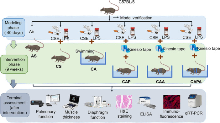
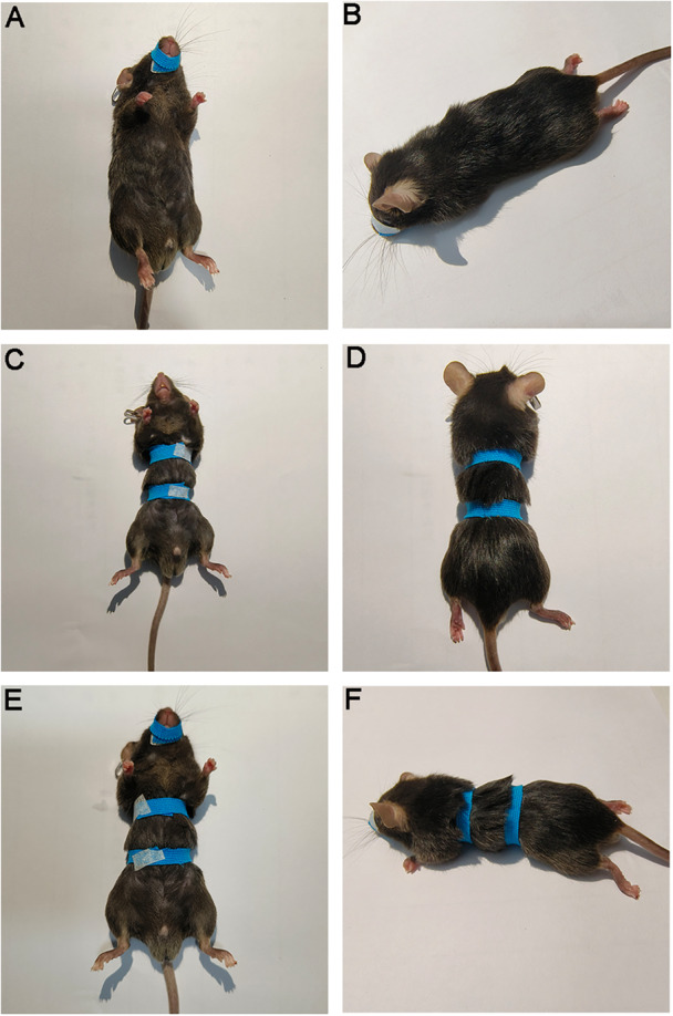
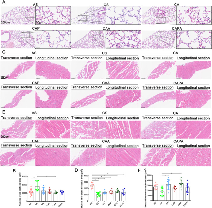
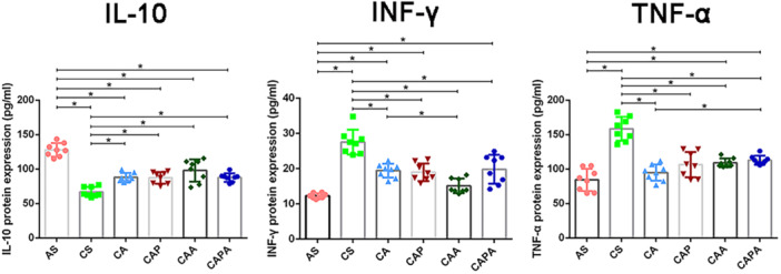
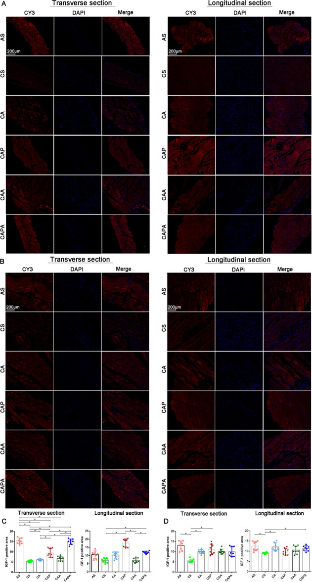
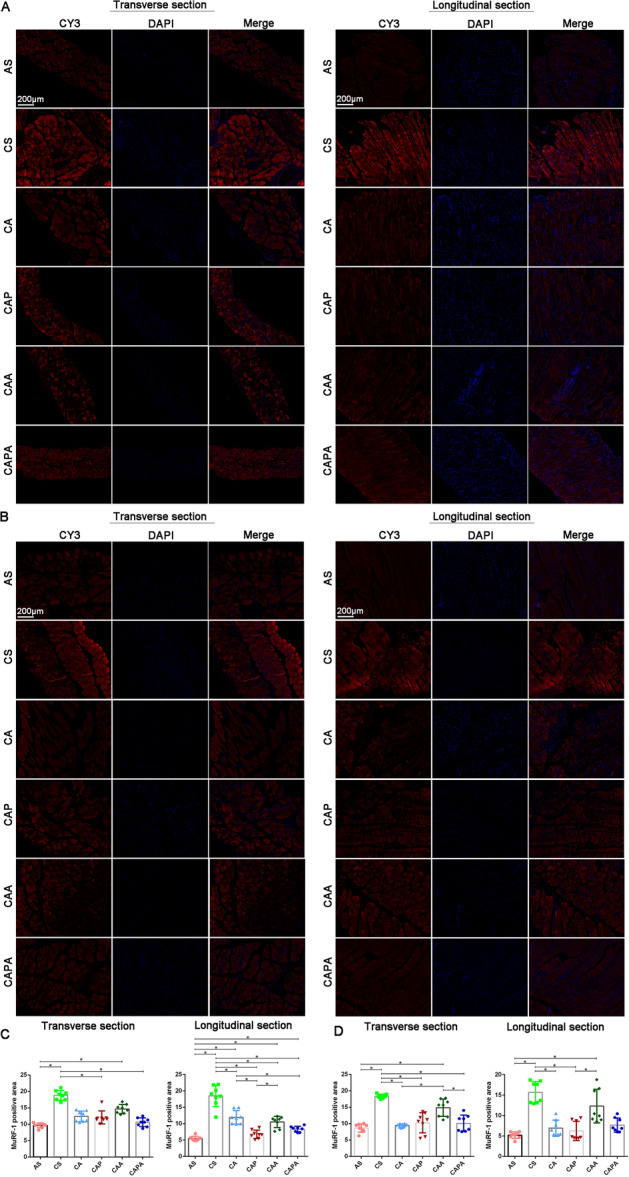
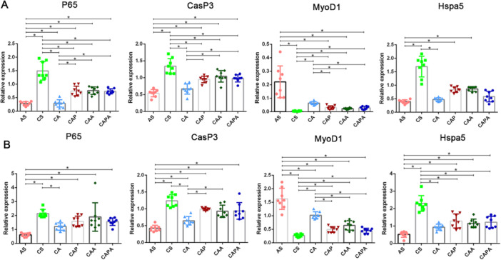
Methods: Forty-eight C57BL/6 mice randomly assigned into six groups to receive interventions mimicking Liuzijue's kinematic features, namely aerobic exercise, pursed-lip breathing and abdominal breathing. Post-intervention, respiratory function, diaphragmatic contractility, and rectus abdominis thickness were assessed. Histological structures of lung tissue, diaphragm, and rectus abdominis were observed using H&E staining. Expression levels of IL-10, INF-γ, and TNF-α in bronchoalveolar lavage fluid, and p65, CasP3, MuRF1, MyoD1, IGF-1, and Hspa5 in the diaphragm and rectus abdominis were measured.

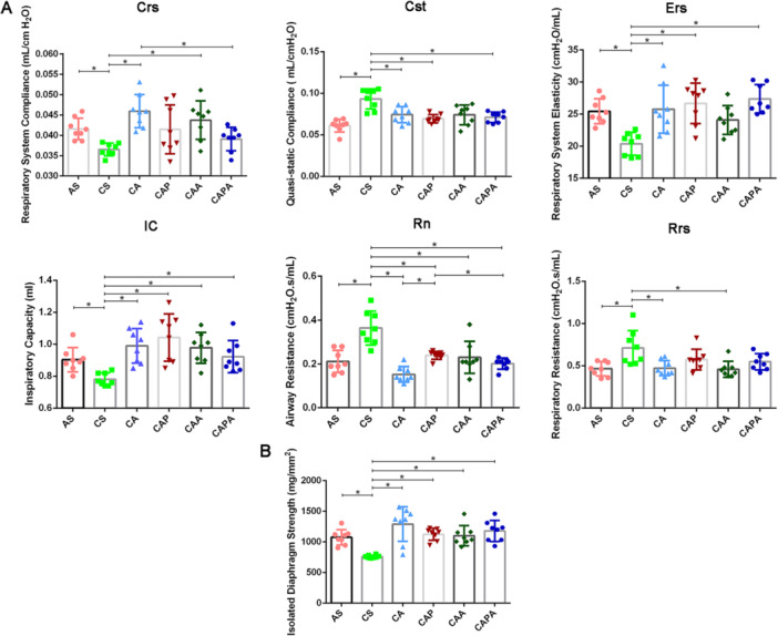
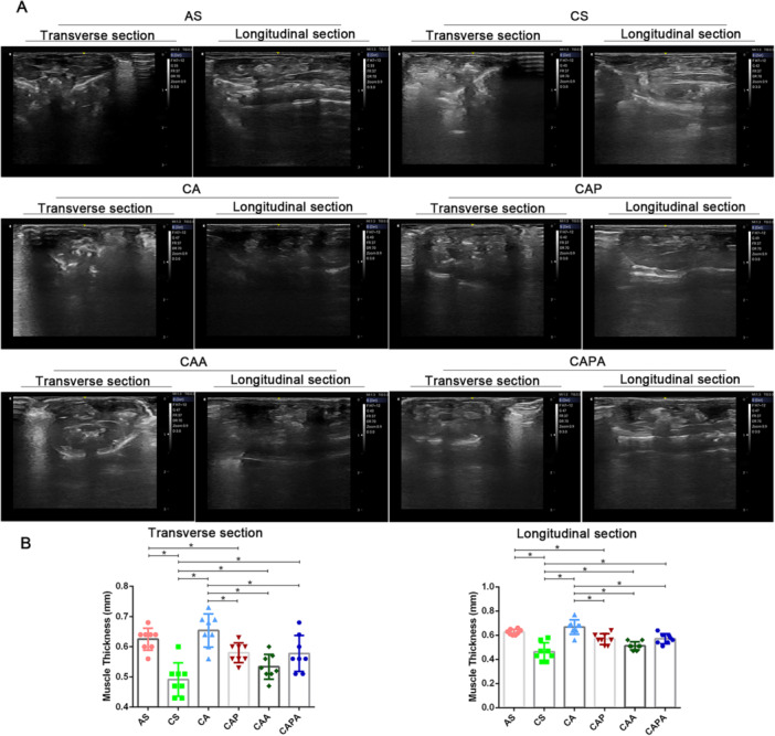
Results: The modeling process impaired respiratory function and diaphragmatic contractility in mice. All four stimulation forms effectively improved pulmonary and diaphragmatic function in COPD mice. The thickness of the rectus abdominis was increased by three specified exercise forms. Despite minimal lung tissue structural changes, swimming and abdominal stimulation improved the structure of the rectus abdominis in COPD mice and airway inflammation levels were inhibited. Lastly, the four stimulations regulated the balance of myoprotein synthesis and degradation in the diaphragm and rectus abdominis, although the intervention effects of the four stimulations did not escalate with the complexity of the methods.

Conclusion: The exercise stimulation paradigms established by simulating the kinematic characteristics of Liuzijue possess the therapeutic effects of Liuzijue in improving respiratory function, demonstrating first evidence that kinematic-based animal models can bridge traditional Qigong and modern mechanism research. However, the development of an animal model for the application of Liuzijue in basic research still warrants further exploration.

Keywords: Liuzijue; animal model; chronic obstructive pulmonary disease; kinematic feature; pulmonary rehabilitation.


Conflict of interest statement

The authors declare no conflicts of interest.

Figures

Figure 1
Figure 1
Schematic diagram of the establishment, application and assessment plan of Liuzijue animal model exploration based on COPD. Mice were randomly assigned into six groups: AS, CS, CA, CAP, CAA, and CAPA (n = 8). Except for the AS group, which served as the control, all other groups were subjected to a 40‐day regimen of cigarette smoke exposure and intra‐airway LPS instillation to establish the COPD mouse model. Subsequently, the CA, CAP, CAA, and CAPA groups underwent a 9‐week intervention involving various forms of exercise. Ultimately, at the end of the 9‐week intervention period, mice were subjected to the following assessments in sequence: pulmonary function testing and rectus abdominis thickness measurements under anesthesia; BALF sampling, tissue collection for diaphragmatic contractility, histopathology, and molecular analyses. AS, blank group; CA, aerobic exercise group; CAA, abdominal muscle stimulation group; CAP, pursed lip breathing group; CAPA, compound stimulation group; CS, model group; CSE, cigarette smoke exposure; ELISA, enzyme‐linked immunosorbent assay; H&E staining, hematoxylin and eosin staining; LPS, lipopolysaccharide; qRT‐PCR, quantitative reverse transcription polymerase chain reaction.
Figure 2
Figure 2
Depiction of kinesiology taping mimicking the kinematic characteristics of Liuzijue Qigong. Panels illustrate the application of kinesiology tape on mice in the CAP group in ventral (A) and dorsal (B) views; the CAA group in ventral (C) and dorsal (D) views; and the CAPA group in ventral (E) and dorsal (F) views. The taping technique is designed to replicate the kinematic features of Liuzijue Qigong, targeting specific muscular engagement and respiratory patterns. AS, blank group; CS, model group; CA, aerobic exercise group; CAP, pursed lip breathing group; CAA, abdominal muscle stimulation group; CAPA, compound stimulation group.
Figure 3
Figure 3
Interventions simulating the kinematic characteristics of Liuzijue Qigong improve pulmonary function and diaphragmatic contractility in COPD mice. Panels display the pulmonary function indicators (A) and diaphragmatic contractility (B) among different groups of mice. Data are expressed as mean ± standard deviation. AS, blank group; CA, aerobic exercise group; CAA, abdominal muscle stimulation group; CAP, pursed lip breathing group; CAPA, compound stimulation group; Crs, respiratory system compliance; CS, model group; Cst, quasi‐static compliance; Ers, respiratory system elasticity; IC, inspiratory capacity; Rn, airway resistance; Rrs, respiratory resistance.
Figure 4
Figure 4
The impact of interventions emulating the kinematic features of Liuzijue on the thickness of the rectus abdominis muscle in COPD mice. Panel (A) displays representative ultrasonic images of the rectus abdominis muscle in mice across different experimental groups; Panel (B) illustrates the comparative analysis of the muscle thickness measured by ultrasound. Data are expressed as mean ± standard deviation. AS, blank group; CS, model group; CA, aerobic exercise group; CAP, pursed lip breathing group; CAA, abdominal muscle stimulation group; CAPA, compound stimulation group.
Figure 5
Figure 5
Morphological changes in respiratory muscles of COPD mice induced by interventions simulating the kinematic characteristics of Liuzijue. Panel (A) presents the structure of lung tissue from different groups of mice, with the scale bar set at 200 μm for the lower magnification and 50 μm for the higher magnification views; Panel (B) compares the cross‐sectional areas of alveoli among the various groups. Panel (C) shows H&E staining of diaphragmatic tissue with a scale bar of 200 μm; Panel (D) illustrates the comparison of cross‐sectional areas of diaphragmatic muscle fibers. Panel (E) depicts H&E staining of the rectus abdominis muscle with a scale bar of 200 μm; Panel (F) presents the comparison of cross‐sectional areas of muscle fibers in the rectus abdominis. Data are expressed as mean ± standard deviation. AS, blank group; CS, model group; CA, aerobic exercise group; CAP, pursed lip breathing group; CAA, abdominal muscle stimulation group; CAPA, compound stimulation group.
Figure 6
Figure 6
Inhibition of airway inflammation in COPD mice by interventions simulating the kinematic characteristics of Liuzijue. Data are expressed as mean ± standard deviation. AS, blank group; CA, aerobic exercise group; CAA, abdominal muscle stimulation group; CAP, pursed lip breathing group; CAPA, compound stimulation group; CS, model group; IL, interleukin; INF, interferon; TNF, tumor necrosis factor.
Figure 7
Figure 7
The promotional effects of interventions derived from the kinematic characteristics of Liuzijue on IGF‐1 expression in respiratory muscles of COPD mice. Panel (A) shows the expression of IGF‐1 in the diaphragm across different groups; Panel (B) presents the expression of IGF‐1 in the rectus abdominis muscle. Panels (C) and (D) depict comparative results of IGF‐1 expression in the diaphragm and rectus abdominis, respectively. Data are expressed as mean ± standard deviation. Bar = 200 μm. AS, blank group; CS, model group; CA, aerobic exercise group; CAP, pursed lip breathing group; CAA, abdominal muscle stimulation group; CAPA, compound stimulation group.
Figure 8
Figure 8
The impact of interventions based on the kinematic characteristics of Liuzijue on MuRF‐1 expression in respiratory muscles of COPD mice. Panel (A) illustrates the MuRF‐1 expression in the diaphragm among various groups; Panel (B) shows the MuRF‐1 expression in the rectus abdominis muscle. Comparative analysis of MuRF‐1 expression in the diaphragm is presented in Panel (C), and in the rectus abdominis in Panel (D). Data are expressed as mean ± standard deviation. Bar = 200 μm. AS, blank group; CS, model group; CA, aerobic exercise group; CAP, pursed lip breathing group; CAA, abdominal muscle stimulation group; CAPA, compound stimulation group.
Figure 9
Figure 9
Effects of interventions simulating the kinematic characteristics of Liuzijue on the expression of inflammatory, autophagic, growth and stress factors in respiratory muscles of COPD mice. Panel (A) presents the comparative transcription levels of P65, CasP3, MyoD1 and Hspa5 in the diaphragm of different groups of mice; Panel (B) presents the comparative transcription levels of P65, CasP3, MyoD1 and Hspa5 in the rectus abdominis of different groups of mice. Data are expressed as mean ± standard deviation. AS, blank group; CS, model group; CA, aerobic exercise group; CAP, pursed lip breathing group; CAA, abdominal muscle stimulation group; CAPA, compound stimulation group.

Similar articles

References

    1. Lozano R., Naghavi M., Foreman K., et al., “Global and Regional Mortality From 235 Causes of Death for 20 Age Groups in 1990 and 2010: A Systematic Analysis for the Global Burden of Disease Study 2010,” The Lancet 380, no. 9859 (2012): 2095–2128. - PMC - PubMed
    1. Ammous O., Feki W., Lotfi T., et al., “Inspiratory Muscle Training, With or Without Concomitant Pulmonary Rehabilitation, for Chronic Obstructive Pulmonary Disease (COPD),” The Cochrane database of Systematic Reviews 1, no. 1 (2023): 013778. - PMC - PubMed
    1. Cavaillès A., Brinchault‐Rabin G., Dixmier A., et al., “Comorbidities of COPD,” European Respiratory Review 22, no. 130 (2013): 454–475. - PMC - PubMed
    1. Xu J., Zeng Q., Li S., Su Q., and Fan H., “Inflammation Mechanism and Research Progress of COPD,” Frontiers in immunology 15 (2024): 1404615. - PMC - PubMed
    1. Pelgrim C. E., Peterson J. D., Gosker H. R., et al., “Psychological Co‐Morbidities in COPD: Targeting Systemic Inflammation, a Benefit for Both?,” European Journal of Pharmacology 842 (2019): 99–110. - PubMed

LinkOut - more resources