. 2025 Jul 23;14(15):1135.
doi: 10.3390/cells14151135.

Loss of C-Terminal Coiled-Coil Domains in SDCCAG8 Impairs Centriolar Satellites and Causes Defective Sperm Flagellum Biogenesis and Male Fertility

Affiliations

Loss of C-Terminal Coiled-Coil Domains in SDCCAG8 Impairs Centriolar Satellites and Causes Defective Sperm Flagellum Biogenesis and Male Fertility

Kecheng Li et al. Cells. .

Abstract

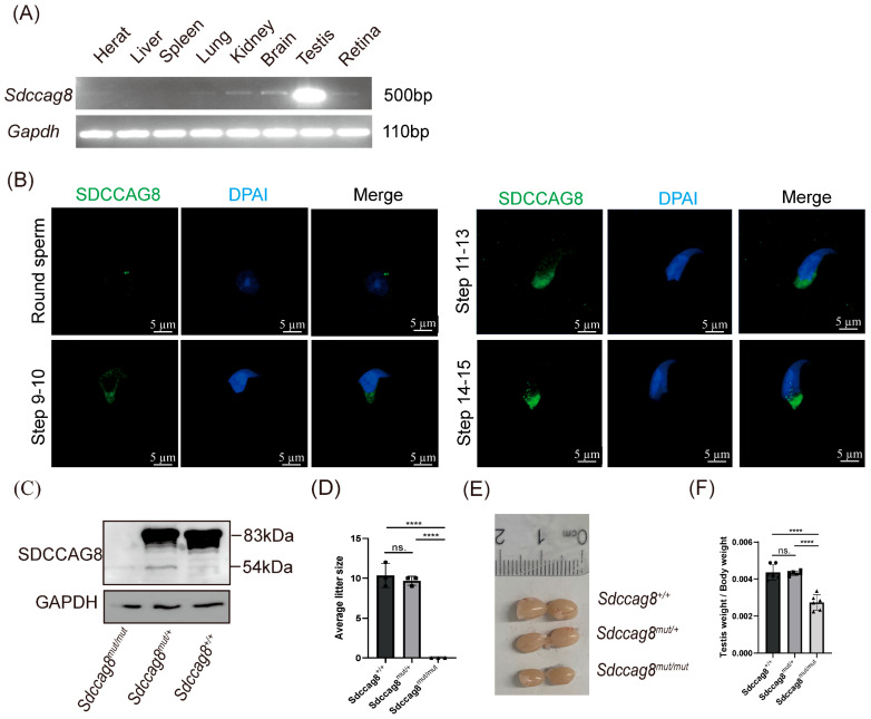
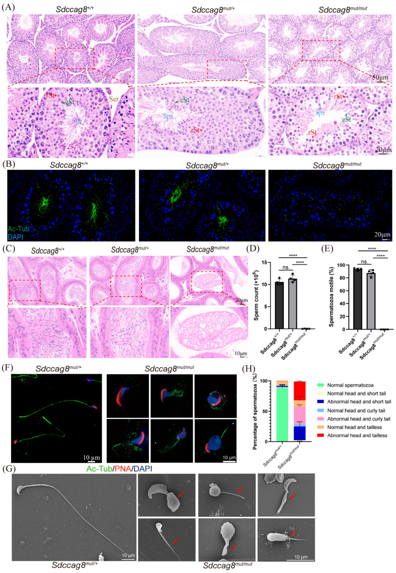
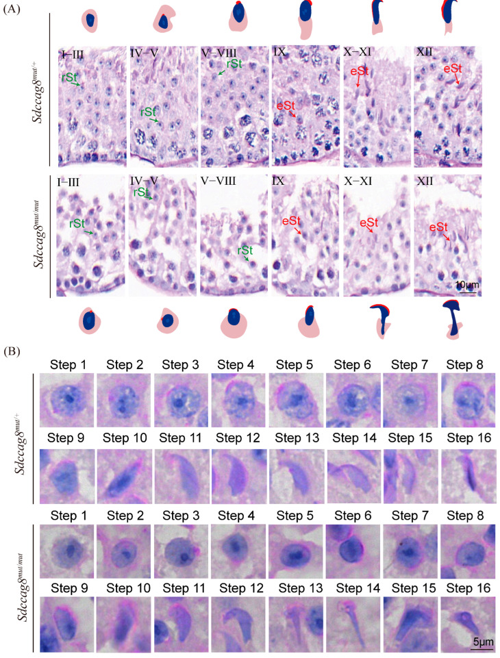
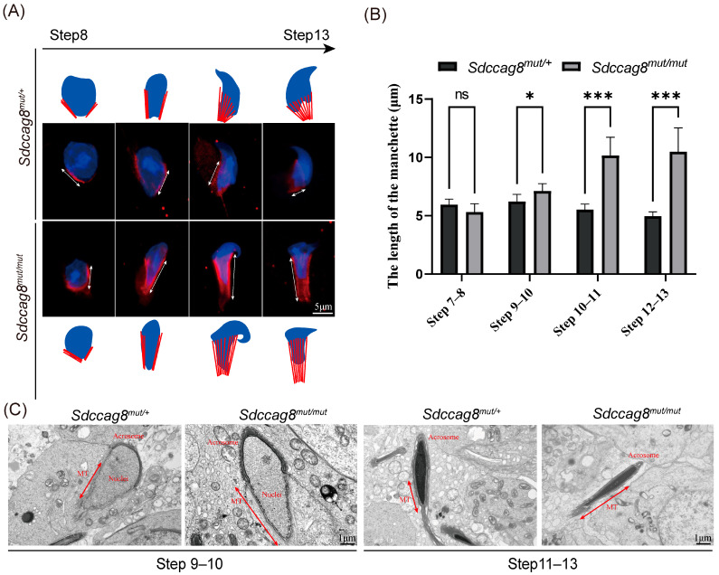
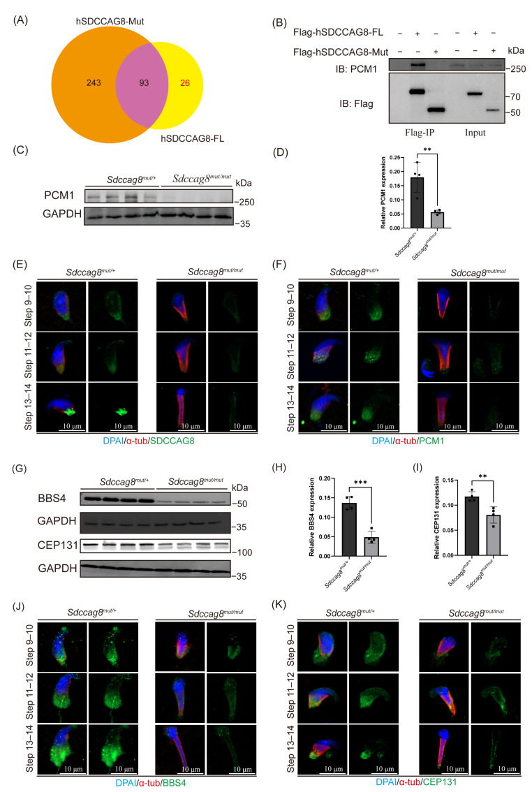
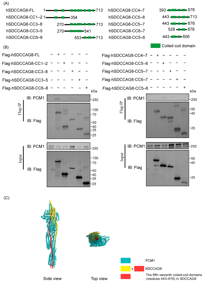
Sperm flagellum defects are tightly associated with male infertility. Centriolar satellites are small multiprotein complexes that recruit satellite proteins to the centrosome and play an essential role in sperm flagellum biogenesis, but the precise mechanisms underlying this role remain unclear. Serologically defined colon cancer autoantigen protein 8 (SDCCAG8), which encodes a protein containing eight coiled-coil (CC) domains, has been associated with syndromic ciliopathies and male infertility. However, its exact role in male infertility remains undefined. Here, we used an Sdccag8 mutant mouse carrying a CC domains 5-8 truncated mutation (c.1351-1352insG p.E451GfsX467) that models the mutation causing Senior-Løken syndrome (c.1339-1340insG p.E447GfsX463) in humans. The homozygous Sdccag8 mutant mice exhibit male infertility characterized by multiple morphological abnormalities of the flagella (MMAF) and dysmorphic structures in the sperm manchette. A mechanistic study revealed that the SDCCAG8 protein is localized to the manchette and centrosomal region and interacts with PCM1, the scaffold protein of centriolar satellites, through its CC domains 5-7. The absence of the CC domains 5-7 in mutant spermatids destabilizes PCM1, which fails to recruit satellite components such as Bardet-Biedl syndrome 4 (BBS4) and centrosomal protein of 131 kDa (CEP131) to satellites, resulting in defective sperm flagellum biogenesis, as BBS4 and CEP131 are essential to flagellum biogenesis. In conclusion, this study reveals the central role of SDCCAG8 in maintaining centriolar satellite integrity during sperm flagellum biogenesis.

Keywords: MMAF; SDCCAG8; centriolar satellites; male infertility; sperm flagellum biogenesis; spermiogenesis.


Conflict of interest statement

The authors declare no conflicts of interest.

Figures

Figure 1
Figure 1
Sdccag8 c. 1351–1352insG mutation leads to male infertility in mice. (A) RT-PCR analysis of Sdccag8 mRNA expression in various tissues from wild-type C57BL/6 adult mice. (B) Immunofluorescence analysis of SDCCAG8 in testicular germ cells from a wild-type C57BL/6 adult mouse. Scale bars: 5 μm. (C) Immunoblot analysis for SDCCAG8 in protein lyases from Sdccag8mut/mut, Sdccag8mut/+, and Sdccag8+/+ testes. (D) The average litter sizes of Sdccag8+/+, Sdccag8mut/+, and Sdccag8mut/mut mice at 3 months. Data are presented as means ± SD. Two-tailed Student’s t test; n.s., not significant; **** p < 0.0001. (E) The general appearance of the testes from Sdccag8+/+, Sdccag8mut/+, and Sdccag8mut/mut. (F) Quantification of the testis weight/body weight ratio in Sdccag8+/+, Sdccag8mut/+, and Sdccag8mut/mut mice (n = 5 mice/group). Data are presented as means ± SD. Two-tailed Student’s t test; n.s., not significant; **** p < 0.0001.
Figure 2
Figure 2
Sdccag8 c. 1351–1352insG mutation leads to MMAF. (A) H&E staining of the testis sections obtained from 2-month-old Sdccag8+/+, Sdccag8mut/+, and Sdccag8mut/mut male mice. eSt, elongating spermatid; rSt, round spermatid; Spz, spermatozoa; Ser, Sertoli cells. Insets are enlarged and shown below the corresponding pictures. Scale bars: 50 μm for origin images and 20 μm for enlarged images. (B) Immunofluorescence analysis of Ac-Tubulin (green) and DAPI (blue) to identify the sperm flagellum in testis sections. Scale bars: 20 μm. (C) H&E staining of the caudal epididymis obtained from 2-month-old Sdccag8+/+, Sdccag8mut/+, and Sdccag8mut/mut male mice. Insets are enlarged and shown below the corresponding pictures. Scale bars: 20 μm for origin images and 10 μm for magnified images. (D) Sperm number obtained from Sdccag8+/+, Sdccag8mut/+, and Sdccag8mut/mut (n = 3). Data are presented as means ± SD. Two-tailed Student’s t test; n.s., not significant; **** p < 0.0001. (E) The percentage of motile spermatozoa from Sdccag8+/+, Sdccag8mut/+, and Sdccag8mut/mut (n = 3). Data are presented as means ± SD. Two-tailed Student’s t test; n.s., not significant; **** p < 0.0001. (F) The immunofluorescence analysis of spermatids from Sdccag8mut/+ and Sdccag8mut/mut mice, the nucleus was stained with DAPI, the sperm flagella were stained with Ac-tubulin (green), the acrosome was stained with PNA lectin (red). Scale bars: 10 μm. (G) Scanning electron microscopy (SEM) analysis of spermatozoa from the epididymides of Sdccag8mut/+ and Sdccag8mut/mut male mice. The red arrows indicate the sperm flagellum with abnormal morphology. Scale bars: 10 μm. (H) The percentages of different spermatozoa observed in Sdccag8mut/+ and Sdccag8mut/mut caudal epididymides. Data are presented as means ± SD.
Figure 3
Figure 3
Spermiogenesis is defective in Sdccag8mut/mut mice. (A) PAS staining of the testis sections obtained from 2-month-old Sdccag8mut/+ and Sdccag8mut/mut male mice. eSt, elongating spermatid; rSt, round spermatid. Scale bars: 10 μm. (B) PAS staining of spermatids at different steps from 2-month-old Sdccag8mut/+ and Sdccag8mut/mut male mice, showing abnormal sperm head shape. Scale bars: 5 μm.
Figure 4
Figure 4
The manchette is abnormally elongated in Sdccag8mut/mut spermatids following step 9. (A) Immunofluorescence analysis of the manchette (double-headed arrows) in Sdccag8mut/+ and Sdccag8mut/mut spermatids with anti-α-tubulin antibody (red). Nuclei were stained with DAPI (blue). The abnormal elongation of manchette in Sdccag8mut/mut spermatids could be recognized following step 9. Scale bars: 5 μm. (B) Statistical analysis of the length of the manchette at different steps. The length of the manchette in spermatids is Sdccag8mut/mut abnormally elongated following step 9. Data are presented as means ± SD. Two-tailed Student’s t test; n.s., not significant; * p < 0.05, *** p < 0.001. (C) Representative TEM micrographs of spermatid heads. The manchette (double-headed arrows) of elongating spermatids (steps 9–13) from Sdccag8mut/mut mice were significantly elongated, twisted, and ectopically placed with an abnormal head shape. Scale bars: 1 μm.
Figure 5
Figure 5
Sdccag8 c.1351–1352insG mutation destabilizes PCM1 in spermatids and disrupts the centriolar satellites’ integrity. (A) Venn diagram showing overlap of proteins between the Flag-hSDCCAG8-FL binding proteins and the Flag-hSDCCAG8-Mut binding proteins. (B) Co-immunoprecipitation analysis of SDCCAG8 and PCM1. (C) Immunoblot analysis of PCM1 and SDCCAG8 in testes of Sdccag8mut/+ and Sdccag8mut/mut. GAPDH was used as a loading control (n = 4). (D) Quantitative results of immunoblot. The protein level of PCM1 was normalized against GAPDH levels. Data are presented as means ± SD. Two-tailed Student’s t test; ** p < 0.01. (E,F) Representative images of spermatids obtained from Sdccag8mut/mut and Sdccag8mut/+ mice testes co-stained with anti-α-tubulin, DAPI (blue), and anti-SDCCAG8 (E), and anti-PCM1 (F) (red). Scale bar: 10 µm. (G) Immunoblot analysis of protein expression level of BBS4 and CEP131 in protein lyases from Sdccag8mut/+ and Sdccag8mut/mut testes. GAPDH served as a loading control (n = 4). (H,I) Quantitative results of immunoblot. Protein levels of BBS4 (H) and CEP131 (I) were normalized against GAPDH levels. Data are presented as means ± SD. Two-tailed Student’s t test; ** p < 0.01, *** p < 0.001. (J,K). Representative images of spermatids obtained from Sdccag8mut/mut and Sdccag8mut/+ mice co-stained with anti-α-tubulin (red), DAPI (blue), and anti-BBS4 (green) (J), and anti-CEP131 (green) (K). Scale bar: 10 µm.
Figure 6
Figure 6
The region consisting of the fifth to the seventh coiled-coil domains in SDCCAG8 (residues 443–676) is essential for the interaction with PCM1. (A) Schematic diagrams of human SDCCAG8-FL, -CC1–2, -CC3–8, -CC3–5, -CC6–8, -CC4–7, -CC5–8, -CC5–7, -CC6–7, -CC7–8. (B) Co-immunoprecipitation analysis of hSDCCAG8 and endogenous PCM1. (C) Binding model of SDCCAG8 and PCM1 was constructed by AlphaFold 3 (https://golgi.sandbox.google.com/about) (accessed on 4 June 2024). The protein complex is visualized by PyMOL. The region of the fifth–seventh coiled-coil domains (residues 443–676) marked by red located in the core area of this protein–protein complex.

Similar articles

References

    1. Ombelet W., Cooke I., Dyer S., Serour G., Devroey P. Infertility and the provision of infertility medical services in developing countries. Hum. Reprod. Update. 2008;14:605–621. doi: 10.1093/humupd/dmn042. - DOI - PMC - PubMed
    1. Boivin J., Bunting L., Collins J.A., Nygren K.G. International estimates of infertility prevalence and treatment-seeking: Potential need and demand for infertility medical care. Hum. Reprod. 2007;22:1506–1512. doi: 10.1093/humrep/dem046. - DOI - PubMed
    1. Tüttelmann F., Ruckert C., Röpke A. Disorders of spermatogenesis: Perspectives for novel genetic diagnostics after 20 years of unchanged routine. Med. Genet. 2018;30:12–20. doi: 10.1007/s11825-018-0181-7. - DOI - PMC - PubMed
    1. Jiao S.Y., Yang Y.H., Chen S.R. Molecular genetics of infertility: Loss-of-function mutations in humans and corresponding knockout/mutated mice. Hum. Reprod. Update. 2021;27:154–189. doi: 10.1093/humupd/dmaa034. - DOI - PubMed
    1. Coutton C., Escoffier J., Martinez G., Arnoult C., Ray P.F. Teratozoospermia: Spotlight on the main genetic actors in the human. Hum. Reprod. Update. 2015;21:455–485. doi: 10.1093/humupd/dmv020. - DOI - PubMed

LinkOut - more resources