. 2025 Jul 24;14(15):1144.
doi: 10.3390/cells14151144.

Granulocyte-Macrophage Colony-Stimulating Factor Inhibition Ameliorates Innate Immune Cell Activation, Inflammation, and Salt-Sensitive Hypertension

Affiliations

Granulocyte-Macrophage Colony-Stimulating Factor Inhibition Ameliorates Innate Immune Cell Activation, Inflammation, and Salt-Sensitive Hypertension

Hannah L Smith et al. Cells. .

Abstract

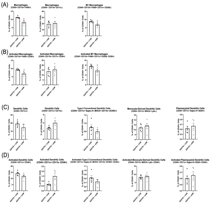
Hypertension (HTN) is a major contributor to global morbidity and manifests in several variants, including salt-sensitive hypertension (SSHTN). SSHTN is defined by an increase in blood pressure (BP) in response to high dietary salt, and is associated with heightened cardiovascular risk, renal damage, and immune system activation. However, the role of granulocyte-macrophage colony-stimulating factor (GM-CSF) has not yet been explored in the context of SSHTN. Previously, we reported that GM-CSF is critical in priming bone marrow-derived (BMD)-macrophages (BMD-Macs) and BMD-dendritic cells (BMD-DCs) to become activated (CD38+) in response to salt. Further exploration revealed these cells differentiated into BMD-M1 Macs, CD38+ BMD-M1 Macs, BMD-type-2 conventional DCs (cDC2s), and CD38+ BMD-cDC2s. Additionally, BMD-monocytes (BMDMs) grown with GM-CSF and injected into SSHTN mice traffic to the kidneys and differentiate into Macs, CD38+ Macs, DCs, and CD38+ DCs. In the current study, we treated SSHTN mice with an anti-GM-CSF antibody (aGM) and found that preventive aGM treatment mitigated BP, prevented renal inflammation, and altered renal immune cells. In mice with established SSHTN, aGM treatment attenuated BP, reduced renal inflammation, and differentially affected renal immune cells. Adoptive transfer of aGM-treated BMDMs into SSHTN mice resulted in decreased renal trafficking. Additionally, aGM treatment of BMD-Macs, CD38+ BMD-M1 Macs, BMD-DCs, and CD38+ BMD-cDC2s led to decreased pro-inflammatory gene expression. These findings suggest that GM-CSF plays a role in SSHTN and may serve as a potential therapeutic target.

Keywords: CD38; granulocyte-macrophage colony-stimulating factor; m1 macrophages; salt-sensitive hypertension; type 2-conventional dendritic cells.


Conflict of interest statement

The authors declare no conflicts of interest.

Figures

Figure 1
Figure 1
Inhibition of granulocyte-macrophage colony-stimulating factor prior to the induction of salt-sensitive hypertension attenuated systolic blood pressure, lowered renal inflammatory gene expression, and mitigated renal function. (A) SBP measurements in CAB-treated and aGM-treated SSHTN mice (n = 4–5). BP was recorded weekly for four weeks and every other day for the final 7 days using the tail-cuff method. (B) Renal expression of inflammation-related genes in CAB-treated and aGM-treated SSHTN mice (n = 4–5). (C) Serum creatinine and urinary creatinine concentrations, FENa, and creatinine clearance in CAB-treated and aGM-treated SSHTN mice (n = 4–5). BP data is presented as the mean ± SD and statistical analyses were performed with an unpaired Student’s t-test, * p < 0.05 vs. SSHTN + CAB. All other data are presented as the mean ± SEM and statistical analyses were performed with an unpaired Student’s t-test, * p < 0.05 vs. SSHTN + CAB.
Figure 2
Figure 2
Preventive anti-granulocyte-macrophage colony-stimulating factor treatment mitigated renal macrophage and dendritic cell differentiation in salt-sensitive hypertension mice. (A) Flow cytometry data assessing Mac populations, as well as (B) their CD38+ counterparts, along with (C) DC populations and (D) their CD38+ counterparts in CAB-treated and aGM-treated SSHTN mice (n = 3–5). Data are presented as mean ± SEM and statistical analyses were performed with an unpaired Student’s t-test, * p < 0.05 vs. SSHTN + CAB.
Figure 3
Figure 3
Treatment with anti-granulocyte-macrophage colony-stimulating factor attenuated systolic blood pressure, reduced renal inflammation, and decreased creatinine clearance in mice with established salt-sensitive hypertension. (A) SBP measurements in CAB-treated and aGM-treated SSHTN mice (n = 4–5). BP was recorded weekly via tail-cuff method. (B) Renal expression of inflammation-related genes in CAB-treated and aGM-treated SSHTN mice (n = 4–5). (C) Serum creatinine and urinary creatinine concentrations, FENa, and creatinine clearance in CAB-treated and aGM-treated SSHTN mice (n = 4–5). BP data is presented as the mean ± SD and statistical analyses were performed with an unpaired Student’s t-test, * p < 0.05 vs. SSHTN + CAB. All other data are presented as the mean ± SEM and statistical analyses were performed with an unpaired Student’s t-test, * p < 0.05 vs. SSHTN + CAB.
Figure 4
Figure 4
Anti-granulocyte-macrophage colony-stimulating factor treatment differentially affected renal Mac and DC subtypes. Renal flow cytometry data assessing (A) Mac populations, as well as their (B) CD38+ counterparts, along with (C) DC populations and (D) their CD38+ counterparts in CAB-treated and aGM-treated SSHTN mice (n = 4–5). Data are presented as the mean ± SEM and statistical analyses were performed with an unpaired Student’s t-test, * p < 0.05 vs. SSHTN + CAB.
Figure 5
Figure 5
Adoptive transfer of anti-granulocyte-macrophage colony-stimulating factor-treated bone marrow-derived monocytes into salt-sensitive hypertension mice reduced macrophage and dendritic cell differentiation. Flow cytometry data assessing the number of (A) CellTracker+ cells grown with GM-CSF or treated with aGM that trafficked to SSHTN kidneys (n = 4). The CellTracker+ cells differentiated into (B) Mac populations, (C) CD38+ Macs, (D) DC populations and (E) CD38+ DCs. Data are presented as the mean ± SEM and statistical analyses were performed with an unpaired Student’s t-test, * p < 0.05 vs. SSHTN + GM.
Figure 6
Figure 6
Anti-granulocyte-macrophage colony-stimulating factor inhibition decreased bone marrow-derived Macrophage differentiation and pro-inflammatory gene expression. Flow cytometry analysis of (A) BMD-Macs and (B) CD38+ BMD-Macs treated with aGM prior to salt (n = 3–5). Cellular expression of inflammation-related genes in BMD-Macs (C) grown with GM-CSF (GM) and treated with salt or BMD-Macs treated with aGM prior to salt. (D) CD38+ M1 Macs were sorted from either BMD-Macs grown with GM-CSF and salt or BMD-Macs treated with aGM and salt to analyze cellular expression of pro-inflammatory genes. Data are presented as the mean ± SEM and statistical analyses were performed with an unpaired Student’s t-test, * p < 0.05 vs. Control + GM or Control + aGM.
Figure 7
Figure 7
Anti-granulocyte-macrophage colony-stimulating factor inhibition decreased bone marrow-derived dendritic cell differentiation and inflammatory gene expression. Flow cytometric analysis of (A) BMD-DCs and (B) CD38+ BMD-DCs treated with aGM prior to salt stimulation (n = 4–5). Cellular expression of inflammation-related genes in BMD-DCs (C) grown with GM-CSF (GM) and treated with salt or BMD-DCs treated with aGM prior to salt. (D) CD38+ cDC2s were sorted from either BMD-DCs grown with GM-CSF and salt or BMD-DCs treated with aGM and salt to analyze cellular expression of inflammatory genes. Data are presented as the mean ± SEM and statistical analyses were performed with an unpaired Student’s t-test, * p < 0.05 vs. Control + GM or Control + aGM; ND = not detectable.

Similar articles

References

    1. Mattson D.L. Immune mechanisms of salt-sensitive hypertension and renal end-organ damage. Nat. Rev. Nephrol. 2019;15:290–300. doi: 10.1038/s41581-019-0121-z. - DOI - PubMed
    1. GBD 2017 Diet Collaborators Health effects of dietary risks in 195 countries, 1990-2017: A systematic analysis for the Global Burden of Disease Study 2017. Lancet. 2019;393:1958–1972. doi: 10.1016/S0140-6736(19)30041-8. - DOI - PMC - PubMed
    1. Elijovich F., Weinberger M.H., Anderson C.A., Appel L.J., Bursztyn M., Cook N.R., Dart R.A., Newton-Cheh C.H., Sacks F.M., Laffer C.L., et al. Salt Sensitivity of Blood Pressure: A Scientific Statement From the American Heart Association. Hypertension. 2016;68:e7–e46. doi: 10.1161/HYP.0000000000000047. - DOI - PubMed
    1. Majid D.S., Prieto M.C., Navar L.G. Salt-Sensitive Hypertension: Perspectives on Intrarenal Mechanisms. Curr. Hypertens. Rev. 2015;11:38–48. doi: 10.2174/1573402111666150530203858. - DOI - PMC - PubMed
    1. Martin S.S., Aday A.W., Almarzooq Z.I., Anderson C.A.M., Arora P., Avery C.L., Baker-Smith C.M., Barone Gibbs B., Beaton A.Z., Boehme A.K., et al. 2024 Heart Disease and Stroke Statistics: A Report of US and Global Data From the American Heart Association. Circulation. 2024;149:e347–e913. doi: 10.1161/CIR.0000000000001209. - DOI - PMC - PubMed

MeSH terms

Substances