. 2025 Jul 25;14(15):1154.
doi: 10.3390/cells14151154.

Molecular Insights into the Superiority of Platelet Lysate over FBS for hASC Expansion and Wound Healing

Affiliations

Molecular Insights into the Superiority of Platelet Lysate over FBS for hASC Expansion and Wound Healing

Sakurako Kunieda et al. Cells. .

Abstract

Human adipose-derived stem cells (hASCs) are widely used in regenerative medicine due to their accessibility and high proliferative capacity. Platelet lysate (PL) has recently emerged as a promising alternative to fetal bovine serum (FBS), offering superior cell expansion potential; however, the molecular basis for its efficacy remains insufficiently elucidated. In this study, we performed RNA sequencing to compare hASCs cultured with PL or FBS, revealing a significant upregulation of genes related to stress response and cell proliferation under PL conditions. These findings were validated by RT-qPCR and supported by functional assays demonstrating enhanced cellular resilience to oxidative and genotoxic stress, reduced doxorubicin-induced senescence, and improved antiapoptotic properties. In a murine wound model, PL-treated wounds showed accelerated healing, characterized by thicker dermis-like tissue formation and increased angiogenesis. Immunohistochemical analysis further revealed elevated expression of chk1, a DNA damage response kinase encoded by CHEK1, which plays a central role in maintaining genomic integrity during stress-induced repair. Collectively, these results highlight PL not only as a viable substitute for FBS in hASC expansion but also as a bioactive supplement that enhances regenerative efficacy by promoting proliferation, stress resistance, and antiaging functions.

Keywords: RNA sequencing; antiaging effects; cell proliferation; human adipose-derived stem cells; platelet lysate; stress resistance.


Conflict of interest statement

The authors declare no conflicts of interest. The funders had no role in the study’s design; in the collection, analyses, or interpretation of data; in the writing of the manuscript; or in the decision to publish the results.

Figures
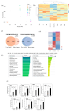
Figure 1
Figure 1
Analysis of RNA sequencing data from hASCs cultured without or with 10% FBS or 3% PL. hASCs were cultured with control (serum free), 10% FBS, and 3% PL-containing DMEM for 48 h. RNA was extracted and used for sequencing. (a) Principal-component analysis (PCA). PCA was performed on log2-transformed TPM values for the 500 most variable genes. Axes denote PC1 (26.7 %) and PC2 (14.8 %). Each point represents one biological replicate (control, n = 2; and FBS and PL, n = 3 per group). (b) Hierarchical clustering heat-map of the 500 most variable genes in the control, FBS, and PL groups. Rows are genes and columns are individual samples; red and blue indicate relative up- and downregulation (row-wise z-scores). Dendrograms were generated with Euclidean distance and Ward’s linkage. (c) Venn diagram of the number of differentially expressed genes (q < 0.05) in the PL and FBS groups compared with the control group. (d) Pearson correlation (heatmap) of the mRNA levels in the control, FBS, and PL groups. The red and blue colors on the y axis represent up- and downregulated genes, respectively. (e) Gene Ontology analysis of upregulated genes (q < 1.0 × 10⁻⁷, β > 4.00). Padj indicates the p-values corrected for multiple testing via the Benjamini–Hochberg method. (f) RT‒qPCR of upregulated mRNAs in hASCs. Relative expression levels of genes related to cell proliferation (FEN1, MT2A, CDC20, and MCM5) and stress resistance (CHEK1, RAD18, and FANCA). The values are expressed as the means ± SDs (n = 3) * p < 0.05, ** p < 0.01 between the indicated groups.
Figure 2
Figure 2
Effect of PL on surviving hASCs treated with 5-FU or H2O2. The cells were cultured with FBS- and PL-containing media and then exposed to 5-FU (a) and H2O2 (b) for 1 h. The values are expressed as the means ± SDs (n = 4) * p < 0.05, ** p < 0.01 between the indicated groups.
Figure 3
Figure 3
Phosphorylation of histone H2A. X (γH2A. X) in hASCs treated with 100 µg/mL 5-FU or 10 µM H2O2. (a) Images of the γH2A. X-positive cells were captured via a fluorescence microscope. Bars = 100 µm. (b) Quantitative analysis of γH2A. X-positive cells. A total of 300 cells were counted under a microscope. The data are expressed as percentages of γH2A. X-positive total cells (n = 3); ** p < 0.01 versus the control.
Figure 4
Figure 4
Effect of PL on the apoptosis of hASCs treated with 5-FU or H2O2. Total apoptosis in hASCs treated with 100 µg/mL 5-FU or 10 µM H2O2 was evaluated via an annexin V assay. (a) Scatchard plots of the results of the apoptosis assay. (b) Quantitative analysis of total apoptotic cells after treatment with 5-FU or H2O2. The values are expressed as the means ± SDs (n = 3). * p < 0.05 and ** p < 0.01 versus the control.
Figure 5
Figure 5
Effect of PL on the doxorubicin-induced aging of hASCs. (a) Fluorescence images of SA-β-gal in hASCs. The cells cultured with 3% PL or 10% FBS were treated with 0.2 µM doxorubicin for 24 h. After the cells were fixed with paraformaldehyde, SA-β-gal was examined and observed via fluoromicroscopy. Bars = 20 μm. (b) Quantitative analysis of the level of SA-β-gal activity normalized to the hASC density. The values are expressed as the means ± SDs of four experiments. ** p < 0.01 versus the control.
Figure 6
Figure 6
Effect of PL on wound healing in vivo. (a) Macroscopic images of control and PL-treated wound areas on days 0, 3, and 7. The yellow broken line indicates the wound margin. Bar = 2.0 mm. (b) Comparison of the average wound area. The wound areas on days 3 and 7 were compared with the original area of the wound on day 0. Data are presented as a percentage of the remaining wound area (each group: n = 5). ** p < 0.01 versus the control.
Figure 7
Figure 7
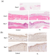
Histological and immunohistochemical analysis of mouse wound healing. (a) Representative HE staining of skin samples from mice treated with or without PL at days 0, 3, and 7. The arrows indicate the position of the capillaries. Bar = 500 µm. (b) Representative immunohistochemical chk1 staining of skin wound samples from mice treated without or with PL at days 3 and 7. Bars = 500 µm.

Similar articles

References

    1. Zuk P.A., Zhu M., Ashjian P., De Ugarte D.A., Huang J.I., Mizuno H., Alfonso Z.C., Fraser J.K., Benhaim P., Hedrick M.H. Human adipose tissue is a source of multipotent stem cells. Mol. Biol. Cell. 2002;13:4279–4295. doi: 10.1091/mbc.e02-02-0105. - DOI - PMC - PubMed
    1. Miana V.V., Prieto González E.A. Adipose tissue stem cells in regenerative medicine. Ecancer Med. Sci. 2018;12:822. doi: 10.3332/ecancer.2018.822. - DOI - PMC - PubMed
    1. Koźlik M., Wójcicki P. The use of stem cells in plastic and reconstructive surgery. Adv. Clin. Exp. Med. 2014;23:1011–1017. doi: 10.17219/acem/37360. - DOI - PubMed
    1. Al-Ghadban S., Bunnell B.A. Adipose tissue-derived stem cells: Immunomodulatory effects and therapeutic potential. Physiology. 2020;35:125–133. doi: 10.1152/physiol.00021.2019. - DOI - PubMed
    1. Mazini L., Rochette L., Admou B., Amal S., Malka G. Hopes and limits of adipose-derived stem cells (ADSCs) and mesenchymal stem cells (MSCs) in wound healing. Int. J. Mol. Sci. 2020;21:1306. doi: 10.3390/ijms21041306. - DOI - PMC - PubMed

LinkOut - more resources