Multicenter Study
. 2025 Jul 26;14(15):1157.
doi: 10.3390/cells14151157.

Pilot Study of [11C]HY-2-15: A Mixed Alpha-Synuclein and Tau PET Radiotracer

Affiliations
Multicenter Study

Pilot Study of [11C]HY-2-15: A Mixed Alpha-Synuclein and Tau PET Radiotracer

Chia-Ju Hsieh et al. Cells. .

Abstract

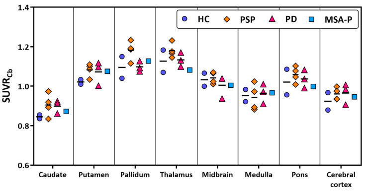
A novel brain positron emission tomography (PET) radioligand, [11C]HY-2-15, has potential for imaging alpha-synuclein aggregations in multiple system atrophy and misfolded tau proteins in tauopathies, based on its high binding affinity in disease brain tissue homogenates. Here, we demonstrate that [3H]HY-2-15 has the capability to bind to aggregated alpha-synuclein in multiple system atrophy brain and tau aggregations in progressive supranuclear palsy and corticobasal degeneration brain tissues via in vitro autoradiography study. A first-in-human pilot multicenter clinical study recruited a total of 10 subjects including healthy controls and patients with Parkinson's disease, multiple system atrophy, or progressive supranuclear palsy. The study revealed that [11C]HY-2-15 has a relatively higher specific uptake in the pallidum and midbrain of patients with progressive supranuclear palsy. Total-body scans performed on the PennPET Explorer showed the radiotracer was cleared by renal excretion. However, the rapid metabolism and low brain uptake resulted in a limited signal of [11C]HY-2-15 in brain.

Keywords: PET; Parkinson’s disease; multiple system atrophy; progressive supranuclear palsy; radioligand; tau; α-synuclein.


Conflict of interest statement

The authors declare no conflicts of interest.

Figures

Figure 1
Figure 1
Autoradiography showing total binding using 10 nM [3H]HY-2-15 (upper panel) and non-specific binding (middle panel; determined with addition of 1 μM cold HY-2-15) of postmortem FFPE brain sections containing the cerebellum from MSA-C, the cingulate cortex from MSA-P, PD, DLB, and HC, the frontal cortex from CBD, and the parietal cortex from PSP cases. Autoradiography color/brightness threshold levels are expressed in counts (0–1000). Per MSA-C, MSA-P, PD, DLB, and HC cases, pS129 α-syn IHC (lower panel) was performed on adjacent sections as pathology reference, while for CBD and PSP, AT8 IHC was carried out. pS129, anti-phospho129 α-syn antibody; AT8, anti-phosphorylated tau antibody. Scale bars: 5 mm. MSA-C, multiple system atrophy—cerebellar subtype; MSA-P, multiple system atrophy—parkinsonian subtype; PD, Parkinson’s disease; DLB, dementia with Lewy bodies; CBD, corticobasal degeneration; PSP, progressive supranuclear palsy; and HC, healthy control.
Figure 2
Figure 2
(A) Parent fraction of [11C]HY-2-15 from arterial blood samples. Time–activity curves of pallidum, midbrain, cerebral cortex, and cerebellar cortex in SUV for (B) HC and (C) PSP. Data is presented as mean ± standard deviation. HC, healthy control; PSP, progressive supranuclear palsy; and SUV, standardized uptake value.
Figure 3
Figure 3
Representative 15–35 min [11C]HY-2-15 SUVR image with a whole brain mask of HC, PSP, PD, and MSA-P in the sagittal and axial views of cerebellum, midbrain, and basal ganglia. HC, healthy control; PSP, progressive supranuclear palsy; PD, Parkinson’s disease; MSA-P, multiple system atrophy—parkinsonian subtype; and SUVR, standardized uptake value ratio.
Figure 4
Figure 4
Scatter plot of brain regional [11C]HY-2-15 SUVRs in HC, PSP, PD, and MSA-P. HC, healthy control; PSP, progressive supranuclear palsy; PD, Parkinson’s disease; MSA-P, multiple system atrophy—parkinsonian subtype; and SUVRCb, standardized uptake value ratio using cerebellar cortex as reference region.
Figure 5
Figure 5
Biodistribution of [11C]HY-2-15. (A) Time–activity curves of %ID in brain and peripheral organs. (B) Total-body SUV image of 7–11 min, 20–30 min, and 80–90 min post [11C]HY-2-15 injection in the two different coronal views of a healthy control subject. SUV, standardized uptake value; and %ID, percent injected dose.

References

    1. Hayes M.T. Parkinson’s disease and parkinsonism. Am. J. Med. 2019;132:802–807. doi: 10.1016/j.amjmed.2019.03.001. - DOI - PubMed
    1. Poewe W., Stankovic I., Halliday G., Meissner W.G., Wenning G.K., Pellecchia M.T., Seppi K., Palma J.-A., Kaufmann H. Multiple system atrophy. Nat. Rev. Dis. Primers. 2022;8:56. doi: 10.1038/s41572-022-00382-6. - DOI - PubMed
    1. Saranza G.M., Whitwell J.L., Kovacs G.G., Lang A.E. Corticobasal degeneration. Int. Rev. Neurobiol. 2019;149:87–136. doi: 10.1016/bs.irn.2019.10.014. - DOI - PubMed
    1. Kovacs G.G., Lukic M.J., Irwin D.J., Arzberger T., Respondek G., Lee E.B., Coughlin D., Giese A., Grossman M., Kurz C. Distribution patterns of tau pathology in progressive supranuclear palsy. Acta Neuropathol. 2020;140:99–119. doi: 10.1007/s00401-020-02158-2. - DOI - PMC - PubMed
    1. Parmera J.B., Oliveira M.C.B.d., Rodrigues R.D., Coutinho A.M. Progressive supranuclear palsy and corticobasal degeneration: Novel clinical concepts and advances in biomarkers. Arq. Neuro-Psiquiatr. 2022;80:126–136. doi: 10.1590/0004-282x-anp-2022-s134. - DOI - PMC - PubMed

Publication types

MeSH terms

LinkOut - more resources