Review
. 2025 Aug 6;14(15):1207.
doi: 10.3390/cells14151207.

Reactive Oxygen Species: A Double-Edged Sword in the Modulation of Cancer Signaling Pathway Dynamics

Affiliations
Review

Reactive Oxygen Species: A Double-Edged Sword in the Modulation of Cancer Signaling Pathway Dynamics

Manisha Nigam et al. Cells. .

Abstract

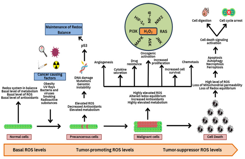
Reactive oxygen species (ROS) are often seen solely as harmful byproducts of oxidative metabolism, yet evidence reveals their paradoxical roles in both promoting and inhibiting cancer progression. Despite advances, precise context-dependent mechanisms by which ROS modulate oncogenic signaling, therapeutic response, and tumor microenvironment dynamics remain unclear. Specifically, the spatial and temporal aspects of ROS regulation (i.e., the distinct effects of mitochondrial versus cytosolic ROS on the PI3K/Akt and NF-κB pathways, and the differential cellular outcomes driven by acute versus chronic ROS exposure) have been underexplored. Additionally, the specific contributions of ROS-generating enzymes, like NOX isoforms and xanthine oxidase, to tumor microenvironment remodeling and immune modulation remain poorly understood. This review synthesizes current findings with a focus on these critical gaps, offering novel mechanistic insights into the dualistic nature of ROS in cancer biology. By systematically integrating data on ROS source-specific functions and redox-sensitive signaling pathways, the complex interplay between ROS concentration, localization, and persistence is elucidated, revealing how these factors dictate the paradoxical support of tumor progression or induction of cancer cell death. Particular attention is given to antioxidant mechanisms, including NRF2-mediated responses, that may undermine the efficacy of ROS-targeted therapies. Recent breakthroughs in redox biosensors (i.e., redox-sensitive fluorescent proteins, HyPer variants, and peroxiredoxin-FRET constructs) enable precise, real-time ROS imaging across subcellular compartments. Translational advances, including redox-modulating drugs and synthetic lethality strategies targeting glutathione or NADPH dependencies, further highlight actionable vulnerabilities. This refined understanding advances the field by highlighting context-specific vulnerabilities in tumor redox biology and guiding more precise therapeutic strategies. Continued research on redox-regulated signaling and its interplay with inflammation and therapy resistance is essential to unravel ROS dynamics in tumors and develop targeted, context-specific interventions harnessing their dual roles.

Keywords: ROS; antioxidants; cancer; oncogenic signaling circuits; oxidative stress mediators; programmed cell death.


Conflict of interest statement

The authors declare no conflicts of interest.

Figures

Figure 1
Figure 1
ROS sources and transformation. The main endogenous and exogenous sources of ROS molecules are depicted in this image, along with the antioxidant mechanisms that catalyze the reduction of various ROS molecules. It also shows the Fenton reaction that produces HO•. NADPH, nicotinamide adenine dinucleotide phosphate (reduced form); NADP+, nicotinamide adenine dinucleotide phosphate (oxidized form); GR, glutathione reductase; GSH, reduced glutathione; GPx, glutathione peroxidase; GSSG, oxidized glutathione; TRX ox, oxidized thioredoxin; Prx, peroxiredoxin; TRX red, reduced thioredoxin; SOD, superoxide dismutase; HO•, hydroxyl radical; O2•−, superoxide ion; H2O2, hydrogen peroxide.
Figure 2
Figure 2
Implications of ROS in cancer. Under homeostatic circumstances, the redox system is in balance in normal cells (green cell), with lower intracellular ROS levels. Factors that boost ROS levels, including obesity, smoking, and radiation exposure, cause DNA damage, genomic instability, and the emergence of cancer-driving mutations in precancerous lesions (red cells). When exposed to factors that increase the risk of developing cancer, tumor suppressor proteins like p53 become active and trigger the transcription of genes related to antioxidant defense. As opposed to this, when repair mechanisms are insufficient, ROS and/or antioxidant levels increase, resulting in an altered redox balance that favors the activation of intracellular oncogenic signaling pathways and maintains the functionality of cellular components in cancer cells (red cells). These routes are often linked to enhanced cytokine release, drug resistance, metastasis, cell survival, and proliferation. Eventually, a breakdown in redox equilibrium brought on by an excessive rise in ROS caused by an overstrained antioxidant system results in apoptosis and necrosis. At this stage, p53 activation brought on by significant cellular damage may result in cell cycle arrest, senescence, and cell death (grey cells), possibly indicating that the response of a variable ROS depends on the tumor’s p53 status and/or oncogenic background. HIF-1α, hypoxia-inducible factor 1-alpha; NF-κB, nuclear factor kappa-light-chain-enhancer of activated B cells; NRF2, nuclear factor erythroid 2-related factor 2; RAS, renin–angiotensin system; JNK, c-Jun N-terminal kinase; ASK, apoptosis signal-regulating kinase; MAPK, mitogen-activated protein kinase; PI3K, phosphoinositide 3-kinase; H2O2, hydrogen peroxide.
Figure 3
Figure 3
ROS-mediated signaling channels. Proteins implicated in cancer-related signaling pathways can be inactivated by ROS (red burst) or activated (green burst) by ROS. ROS stimulate the NF-κB pathway, favoring dimerization of IKK/NEMO and inducing phosphorylation of NF-kB inhibitors (IBs), which are later destroyed by the proteasome. ROS also cause the translocation of NF-κB to the nucleus, causing or inhibiting the transcription of genes involved in cell survival, proliferation, and ROS. Once Keap1 is oxidized, NRF2 is activated, which prevents Keap1 from binding to NRF2, causes NRF2 to relocate to the nucleus, and triggers the transcription of genes that respond to antioxidants. Prolyl-hydroxylases (PDHs), which are essential for joining the p-VHL protein with HIF-1, are also more likely to be oxidized by ROS, leading to their ubiquitination and destruction in the proteasome. In the presence of ROS, TRX oxidizes and separates from ASK. This causes the phosphorylation of MKK, which, in turn, phosphorylates JNK or p38. This facilitates the transcription of genes involved in cell division and proliferation, apoptosis, inflammation, and stress response. As a result of ROS inhibiting SHP-2 phosphatase, TRK receptors continue to be phosphorylated and activate cell signaling, including the RAS-RAF-MEK-ERK pathway, which, in turn, stimulates the transcription of genes that control cell proliferation and differentiation. Lastly, ROS oxidize and inactivate PTEN phosphatase, which encourages the activation of the PI3K/AKT pathway. IGF, insulin-like growth factor; EGF, epidermal growth factor; PDGF, platelet-derived growth factor; TKR, tyrosine kinase receptor; NOX, NADPH oxidase; SOD, superoxide dismutase; H2O2, hydrogen peroxide; ER, endoplasmic reticulum; STAT1/3, signal transducer and activator of transcription 1/3; c-Myc, cellular Myc oncogene; Elk1, Ets-like protein-1; PI3k, phosphoinositide 3-kinase; AKT, protein kinase B; mTOR, mammalian target of rapamycin; PTEN, phosphatase and tensin homolog; RAS, renin–angiotensin system; RAF, rapidly accelerated fibrosarcoma; SHP-2, Src homology 2 domain-containing phosphatase-2; MEK, mitogen-activated protein kinase; ERK, extracellular signal-regulated kinase; MKK, MAP kinase; TRAF6, TNF receptor-associated factor 6; LPS, lipopolysaccharide; TNF-α, tumor necrosis factor-alpha; TNFR, TNF receptor; TLR4, Toll-like receptor 4; TRAF2, TNF receptor-associated factor 2; ASK, apoptosis signal-regulating kinase; JNK, c-Jun N-terminal kinase; IKK-β, IκB kinase-beta; IKK-α, IκB kinase-alpha; NEMO, NF-kappa-B essential modulator; IkB, inhibitor of kappa B; p50, nuclear factor kappa B subunit 1; p65, nuclear factor kappa B subunit 3; PKAc, protein kinase A catalytic subunit; keap 1, Kelch-like ECH-associated protein 1; Cul 3, Cullin-3; NRF2, nuclear factor erythroid 2-related factor 2; P-VHL, von Hippel–Lindau tumor suppressor protein; HIF1-α, hypoxia-inducible factor 1-alpha; PHDs, prolyl hydroxylases; HIF1-β, hypoxia-inducible factor 1-beta; NFAT, nuclear factor of activated T cells; p53, tumor protein p53; Cyclin D1, cyclin D1; HO-1, heme oxygenase-1; GPX1, glutathione peroxidase 1; VEGF, vascular endothelial growth factor; NOX2, NADPH oxidase 2; IL-6, interleukin-6; ΧO, choline oxidase; IL-1ß, interleukin-1 beta; iNOS, inducible nitric oxide synthase; COX2, cyclooxygenase-2; CAT, catalase; CYP, cytochrome P450; TRX, thioredoxin; Bcl-2, B-cell lymphoma 2; Bcl-XL, B-cell lymphoma-extra-large; GSR, glutathione reductase; G6PD, glucose-6-phosphate dehydrogenase; xCT, cystine/glutamate transporter; GCL, glutamate–cysteine ligase; GPX, glutathione peroxidase; PRDX, peroxiredoxin; TXNRD1, thioredoxin reductase 1; CXCR4, C-X-C chemokine receptor type 4.
Figure 4
Figure 4
Thioredoxin (Trx) oxidation and apoptosis signal-regulating kinase 1 (ASK1) activation are caused by an increase in ROS levels in the cytoplasm. ASK1 phosphorylates mitogen-activated protein kinases (ERKs, JNKs, p38 MAPKs), which, in turn, regulate the production of death ligands (FasL), pro-apoptotic proteins (Bak, Bax), anti-apoptotic proteins (Bcl-2, Bcl-xL), and pro-apoptotic proteins (Bcl-2, Bcl-xL). In this way, ERKs encourage cell death by reducing the activity of AKT kinase, which regulates the creation of Bcl-2 and Bcl-xL proteins via NF-κB. The oxidative damage to the mitochondria causes the release of cytochrome c (Cyt. c) into the cytosol, reduction in ATP levels, and dissipation of mitochondrial membrane potential. Caspase-dependent apoptosis is brought on by Cyt. c’s interaction with apoptotic protease activating factor 1 (Apaf-1). Ataxia telangiectasia mutated (ATM) and ataxia telangiectasia and Rad 3-related (ATR) kinases are activated because of an increase in ROS levels in the nucleus, which causes p53-mediated apoptosis and is responsible for the rise in the DNA double-strand breaks marker (H2AX) level. Beclin-1, a pro-autophagic protein, is upregulated by JNK1 in response to the ROS-induced endoplasmic reticulum stress, whereas mTOR kinase, an inhibitor of autophagy, is downregulated by AMPK. Both procedures result in autophagy of the cell. The downregulation of plasma membrane calcium ATPase 1 (PMCA1), a decrease in ATP levels, and an increase in calcium ion levels are all consequences of oxidative damage to the plasma membrane. As a result, cell necrosis happens next. JNK1, c-Jun N-terminal kinase 1; Bcl-2, B-cell lymphoma 2; mTOR, mammalian target of rapamycin; AMPK, AMP-activated protein kinase; ROS, reactive oxygen species; AMP/ATP, AMP/adenosine triphosphate ratio; Bax, Bcl-2-associated X protein; Cyt. c, cytochrome c; Apaf-1, apoptotic protease-activating factor 1; proCSPS-9, pro-caspase-9; CSPS-9, caspase-9; Bcl-xL, B-cell lymphoma-extra-large; ATP, adenosine triphosphate; ΔΨm, mitochondrial membrane potential; CSPS-8, caspase-8; CSPS-3, caspase-3; proCSPS-8, procaspase-8; FasL, Fas ligand; Fas, Fas receptor; PMCA1, plasma membrane calcium-transporting ATPase 1; ICAD, inhibitor of caspase-activated DNase; p53, tumor protein p53; NF-κB, nuclear factor kappa-light-chain-enhancer of activated B cells; ASK-1, apoptosis signal-regulating kinase 1; Trx, thioredoxin; AKT, protein kinase B; ERK, extracellular signal-regulated kinase; p38MAPK, p38 mitogen-activated protein kinase; AP-1, activator protein 1; γ-H2AX, phosphorylated histone H2AX; ATM, ataxia-telangiectasia mutated; ATR, ataxia telangiectasia and Rad3-related.

Similar articles

References

    1. Krieghoff-Henning E., Folkerts J., Penzkofer A., Weg-Remers S. Cancer—An Overview. Med. Monatsschr. Pharm. 2017;40:48–54. - PubMed
    1. Behl T., Kumar A., Vishakha, Sehgal A., Singh S., Sharma N., Yadav S., Rashid S., Ali N., Ahmed A.S., et al. Understanding the Mechanistic Pathways and Clinical Aspects Associated with Protein and Gene Based Biomarkers in Breast Cancer. Int. J. Biol. Macromol. 2023;253:126595. doi: 10.1016/j.ijbiomac.2023.126595. - DOI - PubMed
    1. Sung H., Ferlay J., Siegel R.L., Laversanne M., Soerjomataram I., Jemal A., Bray F. Global Cancer Statistics 2020: GLOBOCAN Estimates of Incidence and Mortality Worldwide for 36 Cancers in 185 Countries. CA. Cancer J. Clin. 2021;71:209–249. doi: 10.3322/caac.21660. - DOI - PubMed
    1. Nigam M., Mishra A.P., Deb V.K., Dimri D.B., Tiwari V., Bungau S.G., Bungau A.F., Radu A.-F. Evaluation of the Association of Chronic Inflammation and Cancer: Insights and Implications. Biomed. Pharmacother. 2023;164:115015. doi: 10.1016/j.biopha.2023.115015. - DOI - PubMed
    1. Khan S.U., Khan M.U., Azhar Ud Din M., Khan I.M., Khan M.I., Bungau S., Hassan S.S. ul Reprogramming Tumor-Associated Macrophages as a Unique Approach to Target Tumor Immunotherapy. Front. Immunol. 2023;14:1–14. doi: 10.3389/fimmu.2023.1166487. - DOI - PMC - PubMed

Substances

LinkOut - more resources