. 2025 Aug 6;14(15):1210.
doi: 10.3390/cells14151210.

Identification of CaVβ1 Isoforms Required for Neuromuscular Junction Formation and Maintenance

Affiliations

Identification of CaVβ1 Isoforms Required for Neuromuscular Junction Formation and Maintenance

Amélie Vergnol et al. Cells. .

Abstract

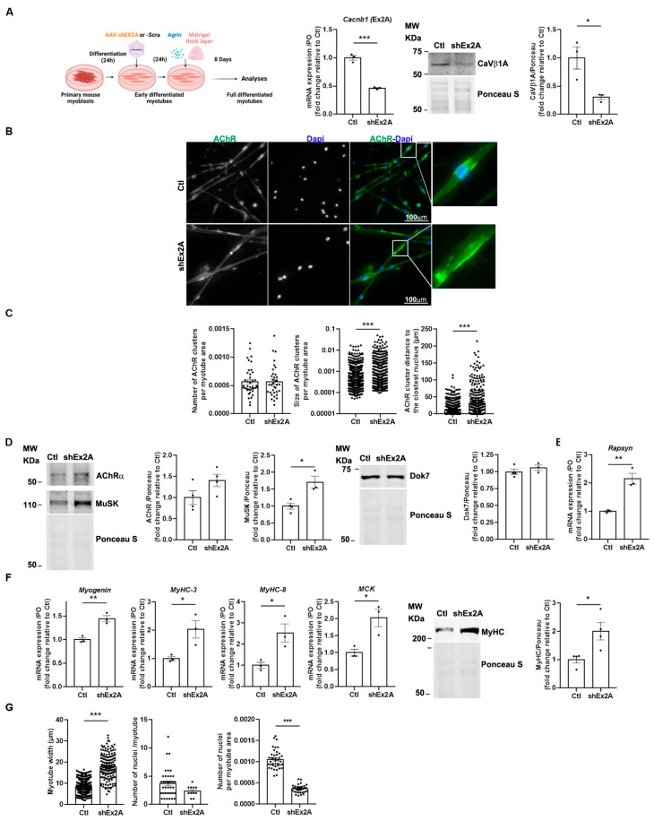
Voltage-gated Ca2+ channels (VGCCs) are regulated by four CaVβ subunits (CaVβ1-CaVβ4), each showing specific expression patterns in excitable cells. While primarily known for regulating VGCC function, CaVβ proteins also have channel-independent roles, including gene expression modulation. Among these, CaVβ1 is expressed in skeletal muscle as multiple isoforms. The adult isoform, CaVβ1D, localizes at the triad and modulates CaV1 activity during Excitation-Contraction Coupling (ECC). In this study, we investigated the lesser-known embryonic/perinatal CaVβ1 isoforms and their roles in neuromuscular junction (NMJ) formation, maturation, and maintenance. We found that CaVβ1 isoform expression is developmentally regulated through differential promoter activation. Specifically, CaVβ1A is expressed in embryonic muscle and reactivated in denervated adult muscle, alongside the known CaVβ1E isoform. Nerve injury in adult muscle triggers a shift in promoter usage, resulting in re-expression of embryonic/perinatal Cacnb1A and Cacnb1E transcripts. Functional analyses using aneural agrin-induced AChR clustering on primary myotubes demonstrated that these isoforms contribute to NMJ formation. Additionally, their expression during early post-natal development is essential for NMJ maturation and long-term maintenance. These findings reveal previously unrecognized roles of CaVβ1 isoforms beyond VGCC regulation, highlighting their significance in neuromuscular system development and homeostasis.

Keywords: CaVβ isoforms; Cacnb1; Long-read sequencing; neuromuscular junctions; promoters; skeletal muscle.


Conflict of interest statement

The authors declare no conflicts of interest.

Figures

Figure 1
Figure 1
Identification of a new CaVβ1 isoform in embryonic muscles. (A) Cacnb1 gene and skeletal muscle transcript variants, Cacnb1A, Cacnb1D, and Cacnb1E, as well as the corresponding protein domains, are represented. Full-length sequencing of the Cacnb1 transcript through Nanopore technology led to the identification of one major isoform per TU. (B) RT-PCR and quantification of the three full-length Cacnb1 variants in innervated (D0) TA muscles or after 3 (D3) and 14 days of denervation. PO was the loading control. (C) Western Blot and quantification of CaVβ1A and CaVβ1D in embryonic (E) and post-natal (P) days. Ponceau S was the loading control. (D) Western Blot and quantification of CaVβ1A and CaVβ1D after 3 (D3) and 14 days of denervation. (E) Western Blot and quantification of CaVβ1A and CaVβ1D in embryonic (E) muscles and adult (P) TA muscles. Ponceau S was the loading control. All data are mean ± SEM (* p < 0.05, ** p < 0.01, and *** p < 0.001). p values were calculated by ordinary one-way ANOVA followed by (BD) Tukey’s and (E) Dunnett’s multiple comparison tests.
Figure 2
Figure 2
Two specific and distinct promoters drive the expression of Cacnb1 isoforms in skeletal muscle. (A) Triplex RT-PCR and quantification of the percentage of exon2A versus exon2B inclusion in embryonic (E) and adult (P) TA muscles. (B) Visualization of epigenetic marks and RNA Polymerase II at Cacnb1 promoters using UCSC Genome Browser. Image displaying the location of exon2B (Ex2B) containing the promoter site 2 (Prom2), exon1 (Ex1) containing the promoter site 1 (Prom1), activating epigenetic marks (H3K3me3, H3K9ac, and H3K27ac), and RNA Polymerase II occupancy at Prom1 and Prom2 in either embryonic (E12.5) muscles (ENCODE project) or adult muscles (Extensor digitorum longus (EDL), Quadriceps, and Soleus). (C) Triplex RT-PCR and quantification of the percentage of exon2A versus exon2B inclusion in innervated and 3-day denervated adult TA muscles (D3). (D) Chromatin immunoprecipitation (ChIP) followed by PCR analysis showing the presence of activating epigenetic marks (H3K4me3 and H3K9ac) and Serine5-phosphorylated RNA Polymerase II (pS5-Pol2) occupancy at Prom1 and Prom2 in innervated and 2-day denervated adult TA/Gas muscles. All data are mean ± SEM (* p < 0.05, ** p < 0.01, *** p < 0.001, and **** p < 0.0001). p values were calculated by ordinary two-way ANOVA followed by Tukey’s multiple comparison test (A), and Sidak’s multiple comparison test (C),or unpaired t-test (D).
Figure 3
Figure 3
Downregulation of embryonic CaVβ1 isoforms induces bigger AChR aggregates and myotube precocious maturation in an in vitro model of highly differentiated myotubes. (A) Schematic representation of the experimental protocol. RT-qPCR of Cacnb1 exon2A (Ex2A), Western Blot, and quantification of CaVβ1A in the control (Ctl) and shEx2-treated myotubes. (B) Immunofluorescence (IF) images of the control or shEx2A-treated myotubes stained with AChRa1 (green) and DAPI (blue). (C) Quantification of AChR cluster number and size per myotube area and distance between AChR clusters and the nearest nuclei. (D) Western Blot and quantification of AChR, MuSK, and Dok7 in the control and shEx2A-treated myotubes. (E) RT-qPCR of Rapsyn in the control and shEx2A-treated myotubes. (F) RT-qPCR of Myogenin, MyHC-3, MyHC-8, and MCK in the control and shEx2A-treated myotubes and Western Blot and quantification of MyHC in the control and shEx2A-treated myotubes. (G) Quantification of the number of nuclei per myotube and number of nuclei per myotube in the control and shEx2A-treated myotubes. All data are mean ± SEM (* p < 0.05, ** p < 0.01, and *** p < 0.001). p values were calculated by an unpaired t-test (AG).
Figure 4
Figure 4
NMJ structural and molecular defects upon downregulation of embryonic CaVβ1 isoforms in TA muscles at post-natal stages. (A) Schematic representation of the experimental protocol. RT-qPCR of embryonic Cacnb1 variants using primer in exon2A in the control and shEx2-treated TA muscles, Western Blot, and quantification of CaVβ1A and CaVβ1D in muscles 3 weeks post-injection. (B) Immunofluorescence (IF) images of the control and shEx2A-treated TA muscles, 3 weeks post-injection, stained with α-bungarotoxin (BTX) for AChR (red), neurofilaments, and Synaptic Vesicle Glycoprotein 2 (NF/SV2) (green) and DAPI (blue). (C) Quantification of NMJ morphology with area of AChR, unoccupied AChR area, overlap between nerve terminals and postsynaptic apparatus, and fragmentation index. (D) Western Blot and quantification of AChRδ, MuSK, and MyHC in the control and shEx2A-treated TA muscles, 3 weeks post-injection. (E) RT-qPCR of Myogenin and Rapsyn in the control and shEx2A-treated muscles. All data are mean ± SEM (* p < 0.05, and *** p < 0.001). p values were calculated by paired (A—Cacnb1 RT-qPCR-, E) and unpaired t-tests (A—CaVβ1A and CaVβ1D WB-, C, D).
Figure 5
Figure 5
NMJ transcriptional and functional defects upon downregulation of embryonic CaVβ1 isoforms in adult TA muscles. (A) Schematic representation of the experimental protocol. RT-qPCR of embryonic Cacnb1 variants using primer in exon2A in the control and shEx2A-treated TA muscles, Western Blot, and quantification of CaVβ1A and CaVβ1D in muscles 12 weeks post-injection. (B) Immunofluorescence (IF) images of the control and shEx2-treated TA muscles, 12 weeks post-injection, stained with α-bungarotoxin (BTX) for AChR (red), neurofilament light chain and Synaptic Vesicle Glycoprotein 2 (NF/SV2) (green), and DAPI (blue). (C) Quantification of NMJ morphology with area of AChR, unoccupied AChR area, overlap between nerve terminals and postsynaptic apparatus, and fragmentation index. (D) Western Blot and quantification of AChRδ, MuSK, and MyHC of Dok7, in the control and shEx2A-treated TA muscles 12 weeks post-injection. (E) RT-qPCR of Myogenin and Rapsyn in the control and shEx2A-treated muscles 12 weeks post-injection. (F) Hematoxylin and eosin (H/E) staining of TA, muscle weight, and cross-sectional area distribution (%) in the control and shEx2A conditions. Bar 50 μm. (G) Indirect (nerve) or direct (muscle) tetanic force measured in the control and shEx2A-treated TA adult muscles, 12 weeks post-injection. CMAP decrement measured by ENMG in the control and shEx2A-treated TA adult muscles, 12 weeks post-injection. All data are mean ± SEM (* p < 0.05, ** p < 0.01, and *** p < 0.001). p values were calculated by paired (A—Cacnb1 RT-qPCR-, E, F, G muscle weight) or unpaired t-tests (A—CaVβ1A and CaVβ1D WB-, C, D) and ordinary two-way ANOVA followed by Sidak’s multiple comparison test (G, CSA distribution).

References

    1. Traoré M., Gentil C., Benedetto C., Hogrel J.-Y., De la Grange P., Cadot B., Benkhelifa-Ziyyat S., Julien L., Lemaitre M., Ferry A., et al. An embryonic CaVβ1 isoform promotes muscle mass maintenance via GDF5 signaling in adult mouse. Sci. Transl. Med. 2019;11 doi: 10.1126/scitranslmed.aaw1131. - DOI - PubMed
    1. Buraei Z., Yang J. Structure and function of the β subunit of voltage-gated Ca2+ channels. Biochim. Et Biophys. Acta (BBA) Biomembr. 2013;1828:1530–1540. doi: 10.1016/j.bbamem.2012.08.028. - DOI - PMC - PubMed
    1. Taylor J., Pereyra A., Zhang T., Messi M.L., Wang Z.-M., Hereñú C., Kuan P.-F., Delbono O. The Cavβ1a subunit regulates gene expression and suppresses myogenin in muscle progenitor cells. J. Cell Biol. 2014;205:829–846. doi: 10.1083/jcb.201403021. - DOI - PMC - PubMed
    1. Vergnol A., Traoré M., Pietri-Rouxel F., Falcone S. New Insights in CaVβ Subunits: Role in the Regulation of Gene Expression and Cellular Homeostasis. Front. Cell Dev. Biol. 2022;10:880441. doi: 10.3389/fcell.2022.880441. - DOI - PMC - PubMed
    1. Chen F., Liu Y., Sugiura Y., Allen P.D., Gregg R.G., Lin W. Neuromuscular synaptic patterning requires the function of skeletal muscle dihydropyridine receptors. Nat. Neurosci. 2011;14:570–577. doi: 10.1038/nn.2792. - DOI - PMC - PubMed

LinkOut - more resources