. 2025 Aug 6;14(15):1211.
doi: 10.3390/cells14151211.

Generation of Individualized, Standardized, and Electrically Synchronized Human Midbrain Organoids

Affiliations

Generation of Individualized, Standardized, and Electrically Synchronized Human Midbrain Organoids

Sanae El Harane et al. Cells. .

Abstract

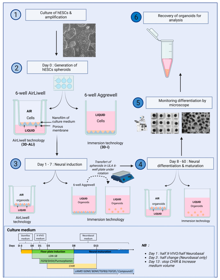
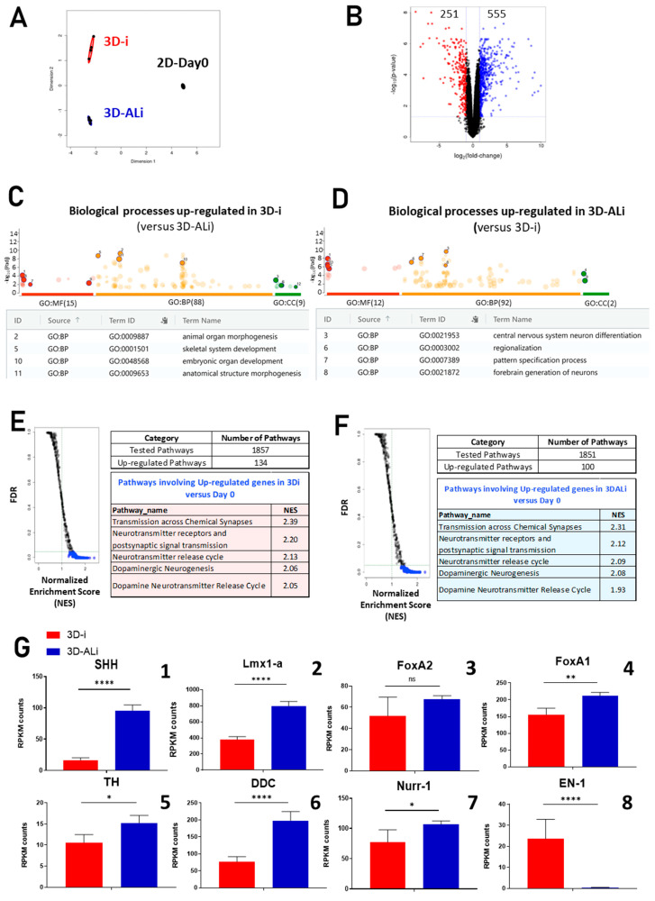
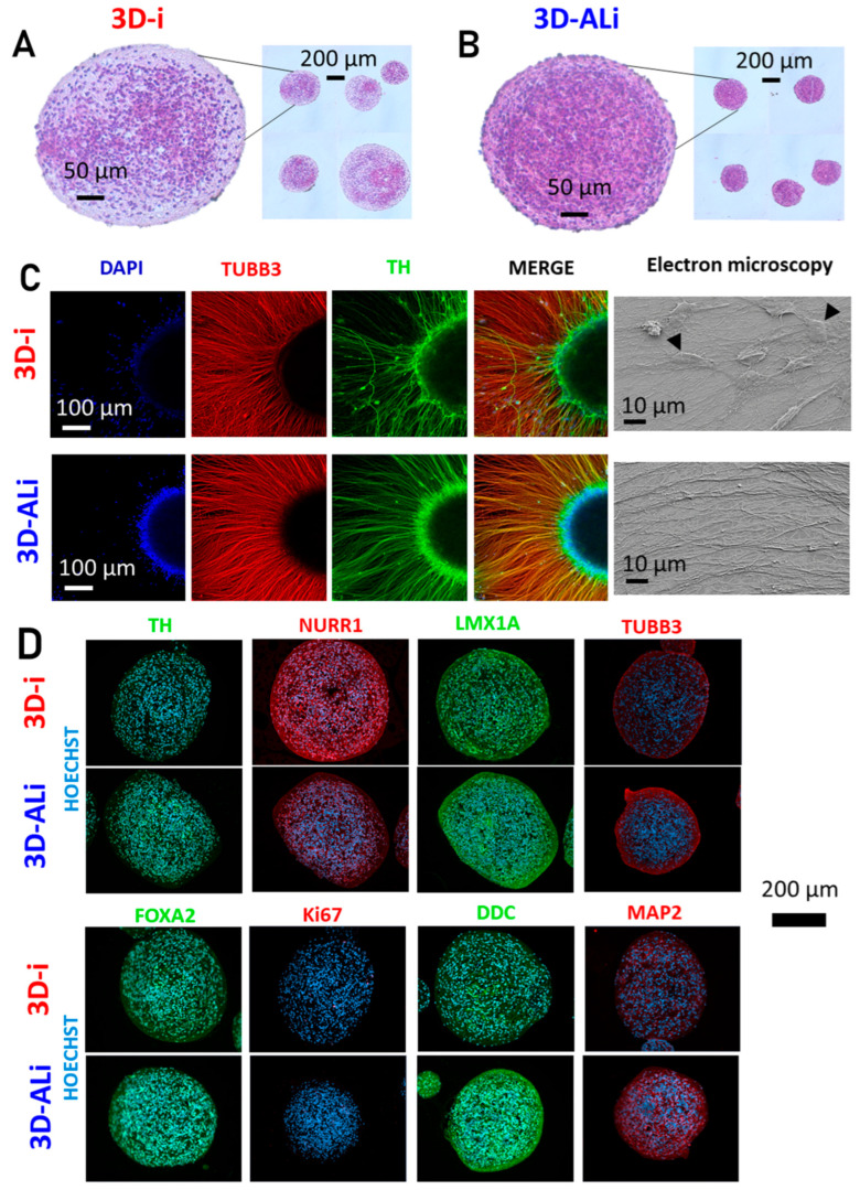
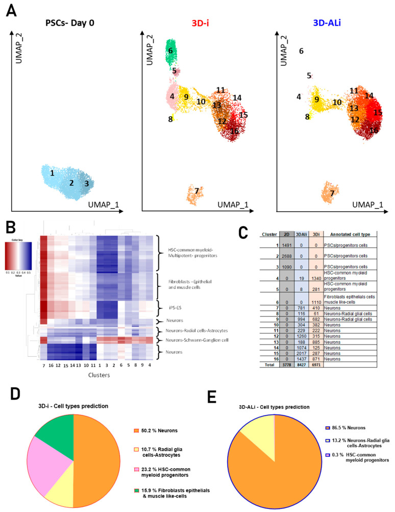
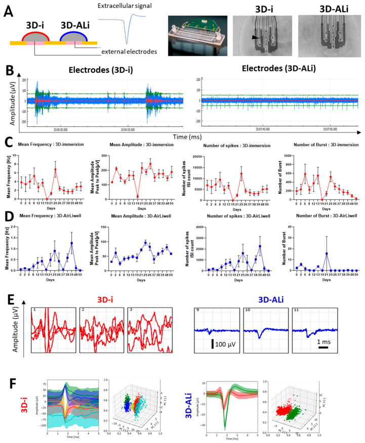
Organoids allow to model healthy and diseased human tissues. and have applications in developmental biology, drug discovery, and cell therapy. Traditionally cultured in immersion/suspension, organoids face issues like lack of standardization, fusion, hypoxia-induced necrosis, continuous agitation, and high media volume requirements. To address these issues, we developed an air-liquid interface (ALi) technology for culturing organoids, termed AirLiwell. It uses non-adhesive microwells for generating and maintaining individualized organoids on an air-liquid interface. This method ensures high standardization, prevents organoid fusion, eliminates the need for agitation, simplifies media changes, reduces media volume, and is compatible with Good Manufacturing Practices. We compared the ALi method to standard immersion culture for midbrain organoids, detailing the process from human pluripotent stem cell (hPSC) culture to organoid maturation and analysis. Air-liquid interface organoids (3D-ALi) showed optimized size and shape standardization. RNA sequencing and immunostaining confirmed neural/dopaminergic specification. Single-cell RNA sequencing revealed that immersion organoids (3D-i) contained 16% fibroblast-like, 23% myeloid-like, and 61% neural cells (49% neurons), whereas 3D-ALi organoids comprised 99% neural cells (86% neurons). Functionally, 3D-ALi organoids showed a striking electrophysiological synchronization, unlike the heterogeneous activity of 3D-i organoids. This standardized organoid platform improves reproducibility and scalability, demonstrated here with midbrain organoids. The use of midbrain organoids is particularly relevant for neuroscience and neurodegenerative diseases, such as Parkinson's disease, due to their high incidence, opening new perspectives in disease modeling and cell therapy. In addition to hPSC-derived organoids, the method's versatility extends to cancer organoids and 3D cultures from primary human cells.

Keywords: 3D cell culture; AirLiwell; Parkinson’s disease; air–liquid interface; cell therapy; electrical recordings; neurospheres; organoids; pluripotent stem cells; spheroids.


Conflict of interest statement

The authors declare no conflicts of interest.

Figures

Figure 1
Figure 1
Highly standardized midbrain organoids generated at air–liquid interface: Illustration of the two 3D cell culture methods: (A) immersion method, (B) AirLiwell method. (C) Illustration of the protocol used to differentiate human pluripotent stem cells (ePSC) into midbrain neurons. Effect of the time culture of dopaminergic differentiation on organoid morphology. (D) Light microscopic photos showing the evolution of organoids during two months (60 days of differentiation) in 3D-ALi compared to 3D-i. The scale bar (left image) is valid for all pictures in panel D and corresponds to 400 μm. (E) Graphs representing the area, roundness, perimeter, circularity, and diameter of midbrain organoids (derived from hPSC, n = 12 organoids quantified for each time point) cultivated in 3D-i and 3D-ALi methods, with statistical significance (p-Value = p) indicated as follows: ns p > 0.05 non-significant, * p < 0.05, ** p < 0.01, *** p < 0.001.
Scheme 1
Scheme 1
Graphical summary of the first part of material and methods regarding the generation and culture of human brain organoids (Created in Biorender. El Harane, S. (2025) https://BioRender.com/frr1adr).
Scheme 2
Scheme 2
Graphical summary of the second part of material and methods regarding the content analysis of organoids (Created in Biorender. El Harane, S. (2025) https://BioRender.com/16yh18m).
Figure 2
Figure 2
Validation of enhanced neural and midbrain specification through bulk RNA sequencing (n = 4; after 6 weeks of differentiation): (A) multidimensional scaling plot; (B) volcano plot depicting regulated genes in 3D-Ali organoids compared to 3D-i organoids; (C) gene ontology analysis results, with a table and representation of up-regulated biological processes, cellular components, and molecular functions in 3D-i organoids and (D) in 3D-ALi organoids; (E) gene set enrichment analysis (GSEA) illustrating the number of regulated pathways compared to pluripotent stem cells at day 0, including the top 15 most up-regulated pathways with their respective normalized enrichment scores (NES) in both immersion and (F) air–liquid interface organoids; (G) graphs displaying gene expression (in RPKM) of genes involved in dopaminergic neurogenesis and late dopaminergic differentiation, with statistical significance (p-Value = p) indicated as follows: ns p >0.05 non-significant, * p < 0.05, ** p < 0.01, **** p < 0.0001 (EDGE R analysis).
Figure 3
Figure 3
Histological comparison reveals enhanced homogeneity in 3D-ALi organoids: (A) Hematoxylin–Eosin colorations of organoid sections from 3D-i organoids, and (B) 3D-ALi organoids at 6 weeks of differentiation. (C) Neuronal maturation of midbrain organoids on poly-ornithine laminin observed in immersion (upper side) and in an air–liquid culture interface (lower side) using immunofluorescence with Anti-TUBB3 (red)/TH (green) and DAPI (blue) staining after 6 weeks of differentiation (including 2 weeks of culture on poly-ornithine laminin). On the right: scanning electron microscopy images showing neuronal networks in both 3D-i and 3D-ALi organoids, and large flattened cells (indicated by black arrows) specifically observed in 3D-i organoids. (D) Immunostaining of organoid sections after 6 weeks of differentiation showing the expression of the following markers: TH (green), NURR1 (red), LMX1A (green), TUBB3 (red), FOXA2 (green), KI67 (red), DDC (green), and MAP2 (red). All nuclei were stained with Hoechst (blue). The respective scale bars are indicated in each image.
Figure 4
Figure 4
3D-ALi organoids contain a high yield of neural cells: (A) UMAPs showing different clusters present in 2D cells at Day 0, in 3D-i organoids, and in 3D-ALi organoids after 6 weeks of differentiation (from left to right); (B) heatmap displaying cell type annotation scores, with each row representing a reference cell subtype and each column representing a cluster from our dataset. Scores are color-coded: positive scores are shown in shades of blue, and negative scores in shades of red. Darker colors indicate stronger associations—either positive or negative—between clusters and cell subtypes. (C) Table showing the number of cells per cluster across all conditions, along with their corresponding annotated cell types. (D) Pie-chart showing the proportion of different cell types found in 3D-i organoids; (E) pie-chart showing the proportion of different cell types found in 3D-ALi organoids. (Orange color: neurons, yellow color: neural progenitors, radial glia cells and astrocytes, pink color: HSC and common myeloid multipotent progenitors, green color: fibroblasts, epithelial and muscle like-cells).
Figure 5
Figure 5
3D-ALi organoids showed a high degree of synchronization of their electrical signal: (A) (From left to right): Schematic illustration of the experimental procedure, the theoretical expected extracellular signal, picture of the microelectrode Array chip used in this experiment, microscopy picture of 3D-i organoids under the electrodes (shown with small black arrow on right), microscopy picture of 3D-ALi organoids under the electrodes; (B) raw recording of the electrical signal in immersion organoids (left) and in air–liquid interface organoids after 4 months of differentiation (right); (C) from left to right, graphs showing the time course (50 days) measurement of mean frequency of electrical activity, mean amplitude, number of spikes, and number of bursts in 3D-i organoids and; (D) in 3D-ALi organoid; (E) pictures of the electrical signal over time and recorded on 3 independent electrodes in 3D-i organoids (on left) and 3D-ALi organoids (on right); (F) principal component analysis graph, showing spike sorting in 3D-i (left) and in 3D-ALi organoids (right). (Each point represents a spike. Each color represents a cluster).

Similar articles

References

    1. Lokai T., Albin B., Qubbaj K., Tiwari A.P., Adhikari P., Yang I.H. A review on current brain organoid technologies from a biomedical engineering perspective. Exp. Neurol. 2023;367:114461. doi: 10.1016/j.expneurol.2023.114461. - DOI - PubMed
    1. Smits L.M., Reinhardt L., Reinhardt P., Glatza M., Monzel A.S., Stanslowsky N., Rosato-Siri M.D., Zanon A., Antony P.M., Bellmann J., et al. Modeling Parkinson’s disease in midbrain-like organoids. npj Park. Dis. 2019;5:5. doi: 10.1038/s41531-019-0078-4. - DOI - PMC - PubMed
    1. Jo J., Xiao Y., Sun A.X., Cukuroglu E., Tran H.D., Goke J., Tan Z.Y., Saw T.Y., Tan C.P., Lokman H., et al. Midbrain-like Organoids from Human Pluripotent Stem Cells Contain Functional Dopaminergic and Neuromelanin-Producing Neurons. Cell Stem Cell. 2016;19:248–257. doi: 10.1016/j.stem.2016.07.005. - DOI - PMC - PubMed
    1. Zhang X.P., Wang X.Y., Wang S.N., Miao C.Y. The generation and properties of human cortical organoids as a disease model for malformations of cortical development. Neural Regen. Res. 2023;18:2119–2126. doi: 10.4103/1673-5374.369100. - DOI - PMC - PubMed
    1. Zhou J.Q., Zeng L.H., Li C.T., He D.H., Zhao H.D., Xu Y.N., Jin Z.T., Gao C. Brain organoids are new tool for drug screening of neurological diseases. Neural Regen. Res. 2023;18:1884–1889. doi: 10.4103/1673-5374.367983. - DOI - PMC - PubMed

Grants and funding

LinkOut - more resources