

Enhanced Response of ZnO Nanorod-Based Flexible MEAs for Recording Ischemia-Induced Neural Activity in Acute Brain Slices
- PMID: 40801712
- PMCID: PMC12348239
- DOI: 10.3390/nano15151173
Enhanced Response of ZnO Nanorod-Based Flexible MEAs for Recording Ischemia-Induced Neural Activity in Acute Brain Slices
Abstract
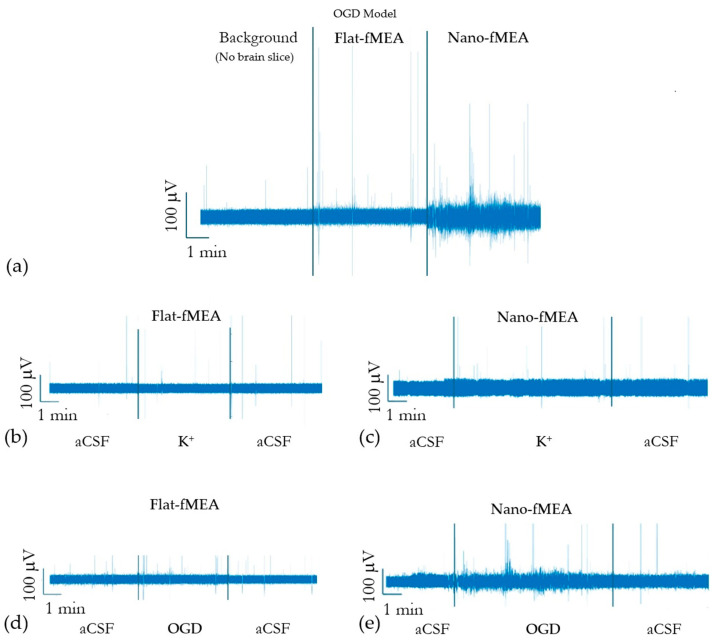
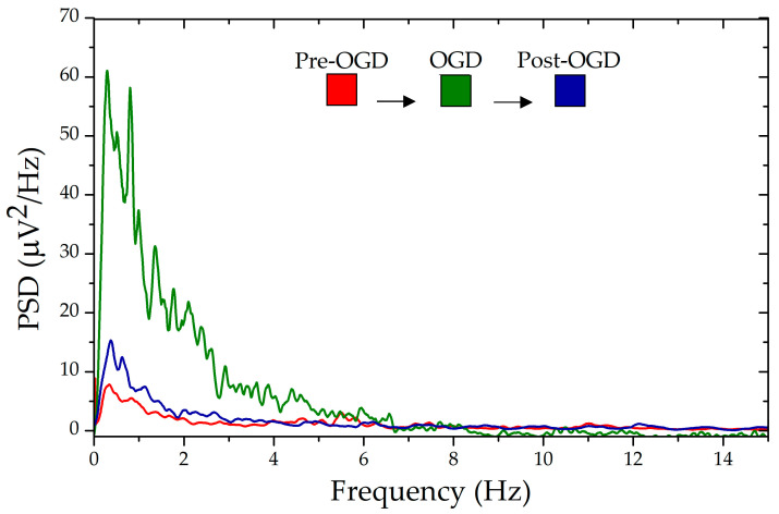
Brain ischemia is a severe condition caused by reduced cerebral blood flow, leading to the disruption of ion gradients in brain tissue. This imbalance triggers spreading depolarizations, which are waves of neuronal and glial depolarization propagating through the gray matter. Microelectrode arrays (MEAs) are essential for real-time monitoring of these electrophysiological processes both in vivo and in vitro, but their sensitivity and signal quality are critical for accurate detection of extracellular brain activity. In this study, we evaluate the performance of a flexible microelectrode array based on gold-coated zinc oxide nanorods (ZnO NRs), referred to as nano-fMEA, specifically for high-fidelity electrophysiological recording under pathological conditions. Acute mouse brain slices were tested under two ischemic models: oxygen-glucose deprivation (OGD) and hyperkalemia. The nano-fMEA demonstrated significant improvements in event detection rates and in capturing subtle fluctuations in neural signals compared to flat fMEAs. This enhanced performance is primarily attributed to an optimized electrode-tissue interface that reduces impedance and improves charge transfer. These features enabled the nano-fMEA to detect weak or transient electrophysiological events more effectively, making it a valuable platform for investigating neural dynamics during metabolic stress. Overall, the results underscore the promise of ZnO NRs in advancing electrophysiological tools for neuroscience research.
Keywords: acute brain slices; cerebral ischemia; micro/nano electrode array; spreading depolarization; zinc oxide nanorods.
Conflict of interest statement
The authors declare no conflict of interest.
Figures

Similar articles
-
Prescription of Controlled Substances: Benefits and Risks.2025 Jul 6. In: StatPearls [Internet]. Treasure Island (FL): StatPearls Publishing; 2025 Jan–. 2025 Jul 6. In: StatPearls [Internet]. Treasure Island (FL): StatPearls Publishing; 2025 Jan–. PMID: 30726003 Free Books & Documents.
-
Short-Term Memory Impairment.2024 Jun 8. In: StatPearls [Internet]. Treasure Island (FL): StatPearls Publishing; 2025 Jan–. 2024 Jun 8. In: StatPearls [Internet]. Treasure Island (FL): StatPearls Publishing; 2025 Jan–. PMID: 31424720 Free Books & Documents.
-
Planar amorphous silicon carbide microelectrode arrays for chronic recording in rat motor cortex.Biomaterials. 2024 Jul;308:122543. doi: 10.1016/j.biomaterials.2024.122543. Epub 2024 Mar 21. Biomaterials. 2024. PMID: 38547834 Free PMC article.
-
Management of urinary stones by experts in stone disease (ESD 2025).Arch Ital Urol Androl. 2025 Jun 30;97(2):14085. doi: 10.4081/aiua.2025.14085. Epub 2025 Jun 30. Arch Ital Urol Androl. 2025. PMID: 40583613 Review.
-
Psychological interventions for adults who have sexually offended or are at risk of offending.Cochrane Database Syst Rev. 2012 Dec 12;12(12):CD007507. doi: 10.1002/14651858.CD007507.pub2. Cochrane Database Syst Rev. 2012. PMID: 23235646 Free PMC article.
References
-
- Serra J., Mateus J.C., Cardoso S., Ventura J., Aguiar P., Leitao D.C. Stress-actuated Flexible Microelectrode Arrays for Activity Recording in 3D Neuronal Cultures. bioRxiv. 2024 doi: 10.1101/2024.12.12.628189. - DOI
-
- Tang X., Shen H., Zhao S., Li N., Liu J. Flexible brain–computer interfaces. Nat. Electron. 2023;6:109–118. doi: 10.1038/s41928-022-00913-9. - DOI
-
- Lacour S.P., Courtine G., Guck J. Materials and technologies for soft implantable neuroprostheses. Nat. Rev. Mater. 2016;1:16063. doi: 10.1038/natrevmats.2016.63. - DOI
Grants and funding
LinkOut - more resources
Full Text Sources
