Case Reports
. 2025 Aug 13;150(1):19.
doi: 10.1007/s00401-025-02899-y.

Biallelic variants in DNAJC7 cause familial amyotrophic lateral sclerosis with the TDP-43 pathology

Affiliations
Case Reports

Biallelic variants in DNAJC7 cause familial amyotrophic lateral sclerosis with the TDP-43 pathology

Toru Yamashita et al. Acta Neuropathol. .

Abstract

Amyotrophic lateral sclerosis (ALS) is a fatal neurodegenerative disorder characterized by the progressive degeneration of motor neurons. ALS pathology primarily involves the failure of protein quality control mechanisms, leading to the accumulation of misfolded proteins, particularly TAR DNA-binding protein 43 (TDP-43). TDP-43 aggregation is a central pathological feature of ALS. Maintaining protein homeostasis is critical and facilitated by heat shock proteins (HSPs), particularly the HSP40 family, which includes co-chaperones such as DNAJC7. Here, we report a family with three siblings affected by ALS who carry a homozygous c.518dupC frameshift variant in DNAJC7, a member of the HSP40 family. All three patients exhibited progressive muscle weakness, limb atrophy, bulbar palsy, and respiratory failure. Pathological examination revealed degeneration of both upper and lower motor neurons, with phosphorylated TDP-43-positive neuronal cytoplasmic inclusions in the frontal and temporal cortices. Immunoblot analysis were consistent with a type B pattern of phosphorylated TDP-43 in the precentral gyrus. Immunohistochemistry and RNA sequencing analyses demonstrated a substantial reduction in DNAJC7 expression at both the protein and RNA levels in affected brain regions. In a TDP-43 cell model, DNAJC7 knockdown impaired the disassembly of TDP-43 following arsenite-induced stress, whereas DNAJC7 overexpression suppressed the assembly and promoted the disassembly of arsenite-induced TDP-43 condensates. Furthermore, in a zebrafish ALS model, dnajc7 knockdown resulted in increased TDP-43 aggregation in motor neurons and reduced survival. To the best of our knowledge, this study provides the first evidence linking biallelic loss-of-function variants in DNAJC7 to familial ALS with TDP-43 pathology.

Keywords: DNAJC7; Amyotrophic lateral sclerosis; Heat shock protein; Live-cell imaging; TDP-43; Zebrafish disease model.


Conflict of interest statement

Declarations. Conflict of interest: The authors declare no potential conflict of interest. Ethical approval: The study was conducted in accordance with the Ethical Guidelines for Human Genome/Gene Analysis Research and the Guidelines for Medical and Health Research Involving Human Subjects, as set forth by the Japanese government. The use of human biological samples was approved by the Ethics Review committees of Okayama University, the National Hospital Organisation Minami-Okayama Medical Centre, Zikei Hospital, and the Tokyo Metropolitan Institute of Medical Science. Written informed consent was obtained from the family members of the deceased.

Figures
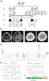
Fig. 1
Fig. 1
Identification of homozygous DNAJC7 variants in three patients with ALS. a The pedigree chart of a family with three ALS patients. The proband is indicated by an arrow. b Brain T1-weighted magnetic resonance images of the proband (IV-5), with red arrows showing characteristic atrophy of the bilateral frontal lobes. Scale bar: 50 mm. c Brain computed tomography images of the proband’s older sister (IV-4), with red arrows indicating atrophy of the bilateral frontal lobes. Scale bar: 50 mm. d Parametric multipoint LOD scores showing significant peaks on chromosomes 5 and 17, suggesting a genetic linkage. e Chromatograms confirming the homozygous c.518dupC variant in DNAJC7 in all three affected individuals. f Schematic representation of the DNAJC7 protein, with the ALS-associated variant located within the tetratricopeptide repeat domain. ALS, amyotrophic lateral sclerosis; LOD, logarithm of odds
Fig. 2
Fig. 2
Phosphorylated TDP-43 pathology, Bunina bodies, and severe loss of the upper and lower motor neurons in the older brother (IV-1) with a homozygous c.518dupC frameshift variant in DNAJC7. a Lateral view of the left hemisphere, showing severe atrophy in the precentral gyrus (asterisks). Arrowhead indicates the top of the precentral gyrus, the width of which is extremely thin. b Severe neuronal loss with gliosis in all cortical layers of the primary motor cortex. Betz cells are nearly completely absent. H&E stain. Scale bar: 200 μm. c Primary motor cortex in a 68-year-old pathologically normal control case. Betz cells are present in layer V, and the structure of the cerebral cortex is spared. H&E stain. Scale bar: 200 μm. d Severe neuronal loss with gliosis in the hypoglossal nucleus. A remaining neuron shows shrinkage of the cell body. H&E stain. Scale bar: 50 μm. e Hypoglossal nucleus in a 51-year-old pathologically normal control case. Neither glial proliferation nor shrinkage of neurons is observed. H&E stain. Scale bar: 50 μm. f Severe degeneration in the pyramidal tract within the medulla oblongata, as visualized by Klüver–Barrera stain. Scale bar: 500 μm. g, h Severe loss of myelin (g) with microglial activation (h) in the lateral tract of the thoracic cord. The dorsal spinocerebellar tract does not exhibit these changes. g Klüver–Barrera stain, h Iba-1 immunohistochemistry. Scale bars: g 1 mm and h 500 μm. i Degeneration in the lateral tract of the lumbar cord, as shown by Klüver–Barrera stain. Scale bar: 1 mm. j Severe loss of anterior horn cells in the lumbar cord, visualized by Klüver–Barrera stain. Scale bar: 500 μm. k Severe gliosis with loss of neurons in the anterior horn of the lumbar cord. H&E stain. Scale bar: 50 μm. l The Spinal anterior horns in a 67-year-old pathologically normal control case. Neither loss of anterior horn cells nor gliosis is present. H&E stain. Scale bar: 50 μm. m Bunina bodies in the lumber anterior horn. H&E stain. Scale bar: 10 μm. n Bunina body in the lumbar anterior horn, identified by Cystatin C immunohistochemistry. Scale bar: 10 μm. o Phosphorylated TDP-43-positive NCIs in the upper layers of the primary motor cortex. Neurites are barely discernible. pS409/410-2 immunohistochemistry. Scale bar: 50 μm. ps Various morphologies of TDP-43-positive NCIs in the lumbar anterior horns. Inclusions include p skein-like, q neurofibrillary change-like, r diffuse granular, and s round structures. pS409/410-2 immunohistochemistry. Scale bars: 10 μm. t Phosphorylated TDP-43-positive glial cytoplasmic inclusion in the pyramidal tract of the medulla oblongata. pS409/410-2 immunohistochemistry. Scale bar: 10 μm. c, e, l Control cases presented here lacked any neurodegenerative changes except for minimal NFTs with Braak stage I. H&E, hematoxylin–eosin; NCI, neuronal cytoplasmic inclusions; NFTs, neurofibrillary tangles; TDP-43, TAR DNA-binding protein of 43 kDa
Fig. 3
Fig. 3
Reduced expression of DNAJC7 protein in the motor cortex of FALS cases with the DNAJC7 variant. a Brain sarkosyl-insoluble fractions from patients with FALS (IV-1, IV-4) and patients with FTLD of various subtypes were subjected to immunoblot analysis using a phosphorylated-TDP43 (Ser409/410) antibody. The immunoblot revealed a distinct pattern of phosphorylated TDP-43, including a prominent band at approximately 50 kDa corresponding to full-length TDP-43, and C-terminal fragments of approximately 23–25 kDa, consistent with the type B pattern previously described [24]. b Representative immunoblot showing DNAJC7 protein levels in motor cortex lysates from two FALS patients (IV-1 and IV-4), two SALS patients, and four controls. β-actin was used as the internal loading control. The asterisk (*) indicates the specific band corresponding to DNAJC7. Notably, DNAJC7 protein was barely detectable in the two FALS cases. c Immunohistochemical staining of brain sections from a patient with FALS (IV-1), a patient with SALS, and a control individual using an anti-DNAJC7 antibody. Scale bar: 50 µm. d Representative images of double -fluorescent staining of SALS brain sections with anti-TDP-43 and anti-DNAJC7 antibodies. Scale bar: 20 µm. SALS, sporadic amyotrophic lateral sclerosis; FALS, familial amyotrophic lateral sclerosis; FTLD, frontotemporal lobar degeneration; TDP-43, TAR DNA-binding protein of 43 kDa
Fig. 4
Fig. 4
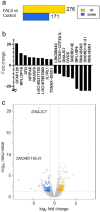
RNA-seq analysis revealed that DNAJC7 expression was decreased in the motor cortex of the FALS cases with the DNAJC7 variant. a Bar graph showing the number of genes that were upregulated or downregulated in patients with FALS (IV-1, IV-4) compared to normal controls. b Waterfall plot depicting the top ten upregulated and downregulated genes in patients with FALS. c Volcano plot illustrating the statistical significance against the fold change for gene expression in patients with FALS. FALS, familial amyotrophic lateral sclerosis; RNA-seq, RNA sequencing
Fig. 5
Fig. 5
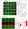
DNAJC7 is required for the disassembly of stress-induced TDP-43 condensates. a Representative living-cell images showing TDP-43ΔNLS-Clover condensates induced by arsenite treatment for 1 h, followed by the removal of stress in U2OS cells treated with control siRNA, or siRNAs targeting HSPA1A, HSPB1, BAG2, and DNAJC7. b Percentage of cells forming TDP-43 condensates after the 1-h arsenite treatment. Data represents means from four independent experiments with individual values shown. c Percentage of cells in which TDP-43 condensates disassemble after 12 h of stress removal. Data represents means from four independent experiments with individual values shown. **p < 0.01 (non-repeated measures analysis of variance and Bonferroni post hoc test). d Representative live-cell images showing TDP-43ΔNLS-Clover condensates induced by arsenite treatment for 1 h, followed by stress removal in U2OS cells transduced with DNAJC7-mCherry lentivirus to increase DNAJC7 levels. e Percentage of cells forming TDP-43 condensates after 1-h arsenite treatment. Data are shown as mean ± SD from two independent experiments. *p < 0.05 (two-tailed unpaired Student’s t-test). f Percentage of cells recovering and disassembling TDP-43 condensates after 12 h of stress removal. Data are shown as mean ± SD from two independent experiments. *p < 0.05 (two-tailed unpaired Student’s t-test)
Fig. 6
Fig. 6
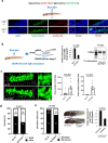
Knockdown of dnajc7 leads to impaired neurodevelopment through accelerated TDP-43 aggregation. a Schematic representation and representative image of optogenetically modified transgenic zebrafish Tg[mnr2b-hs:opTDP-43h] Tg[mnr2b-hs:EGFP-TDP-43z]. The opTDP-43h construct encodes a human TDP-43 anchoring optic module, cryptochrome (CRY2), and RFP. Under basal conditions, the red signal is weakly expressed in the nucleus. After blue light illumination for 48–96 h, a clear aggregation of TDP-43 is observed at perinuclear sites. EGFP-TDP-43z encodes zebrafish TDP-43, which is recruited to and aggregated at the perinuclear site upon light-induced aggregation of opTDP-43 (yellow foci indicated by white arrows). b (Left) Schematic representation of CRISPR-Cas9-mediated dnajc7 knockout in zebrafish. (Middle) Quantitative reverse transcription PCR analysis of dnajc7 expression levels in sg ctr and sg dnajc7 injected embryos at 6 dpf (days post-fertilization). (Right) Agarose gel image showing the T7 endonuclease I assay results for the sgRNAs targeting dnajc7, confirming successful knockout. c (Left) Representative microscopic images showing TDP-43 aggregates in sg ctr and sg dnajc7 injected ALS zebrafish. (Right) Quantification of pixel count for TDP-43 aggregates in sg ctr (7 fields) and sg dnajc7 (8 fields) injected ALS zebrafish at 6 dpf. Data are presented as mean ± SD from one of three independent experiments. Statistical differences were calculated using the Mann–Whitney U-test. d Fraction of dead embryos in sg ctr and sg dnajc7 injected ALS zebrafish at 6 dpf. Data show the number of dead or survived embryos from one of three independent experiments. Differences were calculated using Fisher’s exact test. e (Left) Fraction of embryos with failed SB inflation in sg ctr and sg dnajc7 injected ALS zebrafish at 6 dpf. (Middle) Representative images of SB in the embryos. (Right) Quantitative reverse transcription PCR analysis of dnajc7 expression in the survived sg dnajc7 injected embryos at 6dpf. Embryos with SB inflation failure showed significantly lower dnajc7 mRNA expression than those with normal SB inflation. Data show mean ± SD from one of three independent experiments. Statistical differences were calculated using Fisher's exact test or Mann–Whitney U-test. SB, swim bladder; ALS, amyotrophic lateral sclerosis; TDP-43, TAR DNA-binding protein of 43 kDa

Similar articles

References

    1. Aoki M, Ogasawara M, Matsubara Y, Narisawa K, Nakamura S, Itoyama Y et al (1993) Mild ALS in Japan associated with novel SOD mutation. Nat Genet 5:323–324. 10.1038/ng1293-323 - PubMed
    1. Arai T, Hasegawa M, Akiyama H, Ikeda K, Nonaka T, Mori H et al (2006) TDP-43 is a component of ubiquitin-positive tau-negative inclusions in frontotemporal lobar degeneration and amyotrophic lateral sclerosis. Biochem Biophys Res Commun 351:602–611. 10.1016/j.bbrc.2006.10.093 - PubMed
    1. Arseni D, Hasegawa M, Murzin AG, Kametani F, Arai M, Yoshida M et al (2022) Structure of pathological TDP-43 filaments from ALS with FTLD. Nature 601:139–143. 10.1038/s41586-021-04199-3 - PMC - PubMed
    1. Arseni D, Nonaka T, Jacobsen MH, Murzin AG, Cracco L, Peak-Chew SY et al (2024) Heteromeric amyloid filaments of ANXA11 and TDP-43 in FTLD-TDP type C. Nature 634:662–668. 10.1038/s41586-024-08024-5 - PMC - PubMed
    1. Asakawa K, Handa H, Kawakami K (2020) Optogenetic modulation of TDP-43 oligomerization accelerates ALS-related pathologies in the spinal motor neurons. Nat Commun 11:1004. 10.1038/s41467-020-14815-x - PMC - PubMed

Publication types

LinkOut - more resources