. 2025 Aug 13;82(1):308.
doi: 10.1007/s00018-025-05846-y.

KRAS4B is required for placental development

Affiliations

KRAS4B is required for placental development

Marie-Albane Minati et al. Cell Mol Life Sci. .

Abstract

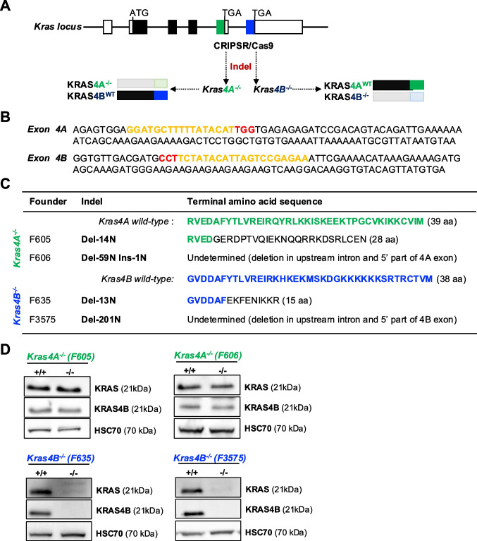
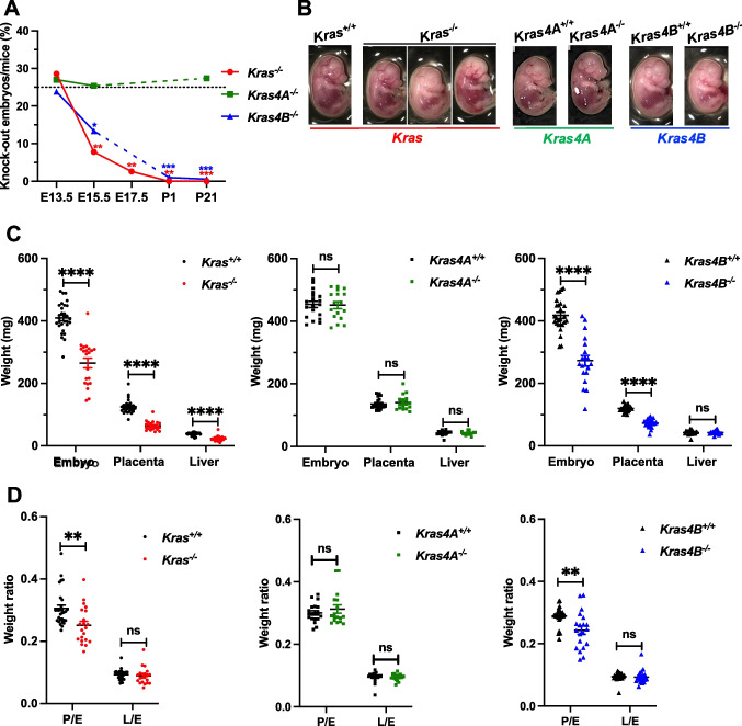
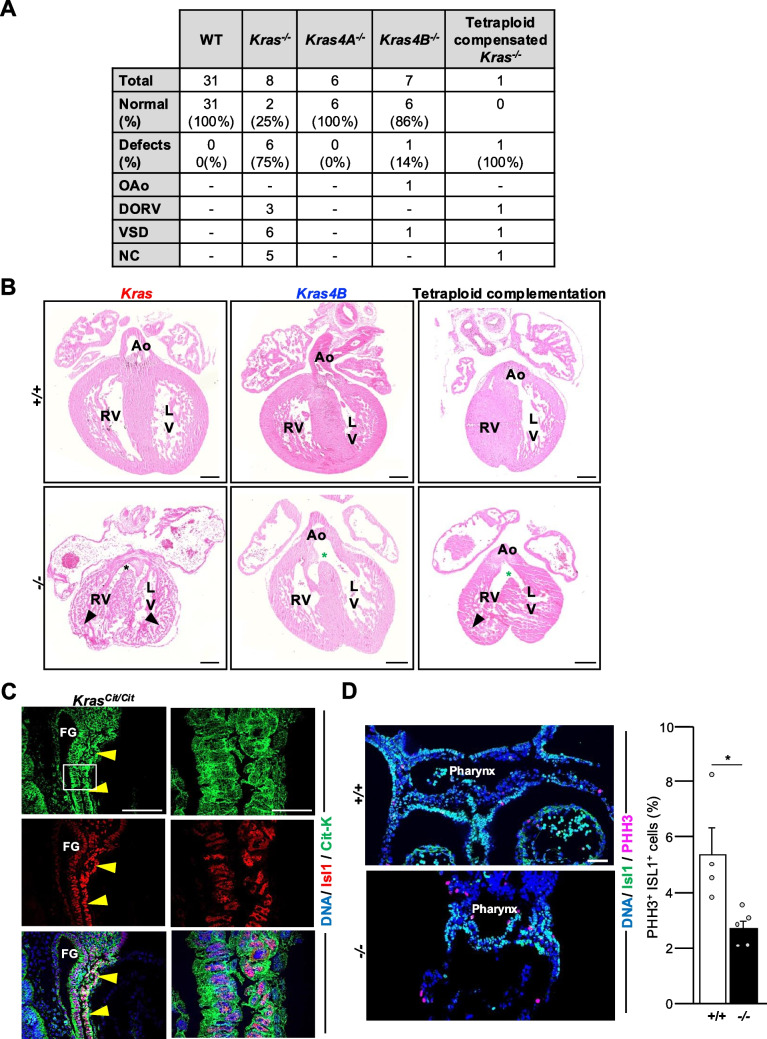
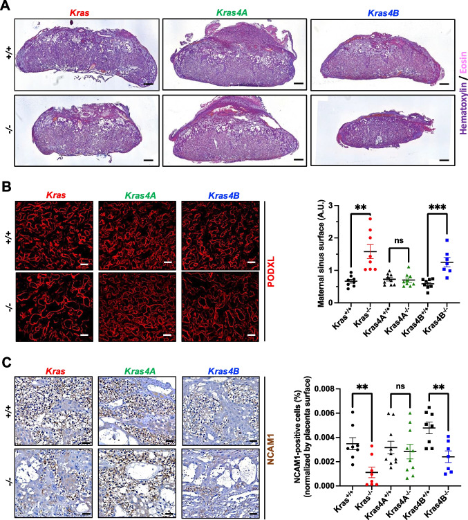
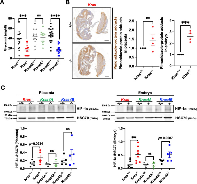
Beyond its well-established role in cancer, KRAS is also crucial for embryogenesis, as its absence leads to embryonic lethality. However, the precise mechanisms underlying the developmental functions of KRAS, as well as the respective roles of its two splicing isoforms, KRAS4A and KRAS4B, remain incompletely characterized. To address these issues, we generated Kras4A knock-out (Kras4A-/-) and Kras4B-/- mouse models using CRISPR/Cas9 technology, and compared their phenotypes to those of a Kras-/- model, in which both isoforms are simultaneously inactivated. We observed that Kras-/- and Kras4B-/- embryos show a lethality that starts around E13.5, while Kras4A-/- embryos develop normally, with no detectable abnormalities. In contrast, Kras-/- embryos displayed a dual phenotype affecting both the heart and placenta, whereas Kras4B-/- embryos exhibited only the placental phenotype. The cardiac phenotype was complex, combining ventricular non-compaction, ventricular septal defects, double outlet right ventricle, and overriding aorta, likely resulting from impaired cardiac precursor proliferation. The placental phenotype was characterized by reduced placental size, and a marked decrease in glycogen trophoblast cells, correlating with hypoglycemia and hypoxia in Kras-/- and Kras4B-/- embryos. Thus, our findings confirm the predominant role of KRAS4B in KRAS-mediated developmental functions, but also suggest hidden functions of KRAS4A. Importantly, this study is the first to identify KRAS as a key regulator of a specific cell differentiation process and to characterize the biological defects caused by its loss.

Keywords: Development; Heart; KRAS4A; KRAS4B; Placenta.


Conflict of interest statement

Declarations. Conflict of interest: The authors declare that the research was conducted in the absence of any commercial or financial relationships that could be construed as a potential conflict of interest.

Figures

Fig. 1
Fig. 1
Generation of the Kras4A−/− and Kras4B−/− mouse models. (A) Scheme depicting the generation of the Kras4A−/− and Kras4B−/− models using CRISPR/Cas9. Genomic structure of the Kras locus is represented by white (non-coding sequences) and black boxes (coding-sequences). Exons 4A and 4B correspond to the green and blue boxes, respectively. The strategy used aims to insert or delete (indel) nucleotides in exons 4 A and 4B by non-homologous DNA end-joining following Cas9 cleavage. (B) Nucleotide sequences of exons 4 A and 4B, with the sequences corresponding to the single guide RNA used (yellow), and the PAM motifs (red). (C) Characteristics of the founders used to generate the Kras4A−/− and Kras4B−/− models. The codes (F…) correspond to the founder mice (2 founders per model). Indel indicates the number of nucleotides (N) inserted (Ins) and/or deleted (Del) in exons 4 A or 4B for each model. Terminal amino acid sequence indicates the single-letter amino acid sequence encoded by exons 4 A (green) and 4B (blue) from the wild-type allele, as well as the aa sequences predicted following the presence of indels in the different models. (D) Western blotting performed on liver extracts from embryos descending from the two founders (F605 and F606) of the Kras4A−/− colony and the two founders (F635 and F3575) of the Kras4B−/− colony. Due to the presumably lower expression of KRAS4A, no decreased KRAS expression (detected with an antibody recognizing both isoforms) is seen in Kras4A−/− embryos. KRAS4B expression (detected with an antibody specifically recognizing KRAS4B) is not affected in Kras4−/− mice. In Kras4B−/− embryos, KRAS4B expression is completely lost, while low KRAS4A expression is detected using the antibody recognizing both isoforms. Heat shock cognate protein 70 (HSC70) was used as loading control
Fig. 2
Fig. 2
Kras−/− and Kras4B−/− mice die in utero and have an impaired growth. (A) Percentage of Kras−/− (red line), Kras4A−/− (green line) and Kras4B−/− (blue line) mice as a function of the embryonic (E) or postnatal (P) stage. Black dot line represents the expected Mendelian percentage (25%) of knock-out (KO) embryos/mice. (B) Pictures showing the macroscopic phenotype of E15.5 embryos. Kras−/− embryos are frequently smaller and some of them present a paleness (embryo in the middle) and haemorrhagic signs (embryo on the right). Kras4A−/− embryos present a normal macroscopic phenotype whereas Kras4B−/− embryos are also frequently smaller. (C) Graphs showing the weight of E15.5 embryo, placenta and liver in the Kras−/−, Kras4A−/− and Kras4B−/− models and their control (+/+) counterparts. (D) Graphs showing the placenta/embryo (P/E) and liver/embryo (L/E) weight ratios at E15.5 for the Kras−/−, Kras4A−/− and Kras4B−/− embryos and their control (+/+) counterparts
Fig. 3
Fig. 3
Kras−/− and Kras4B−/− embryos, but not Kras4A−/− embryos, develop congenital heart defects. (A) Cardiac defects scored after analysis of histological sections at E15.5 Kras−/−, Kras4A−/−, Kras4B−/−, tetraploid complemented Kras−/−, and Kras+/+ embryos. OAo, overriding aorta; DORV, double outlet right ventricle; VSD, ventricular septal defect; NC, non-compaction. (B) Eosin-stained sections of E15.5 hearts revealing that Kras−/−, Kras4B−/−, and tetraploid complemented Kras−/− embryos display congenital heart defects including membraneous ventricular septal defect (asterisk) associated with an overriding aorta (green asterisk) and ventricular non-compaction (arrowhead). Ao, aorta; LV, left ventricle; RV, right ventricle. Scale bars: 200 µm. (C) Immunofluorescent labelling for Citrine-Kras (Cit-K, using a GFP antibody) and Isl1 (for the cardiac progenitors) performed on E10.5 KrasCit/Cit embryos. The white square corresponds to the magnified regions on the right. FG, foregut. Yellow arrow heads: cardiac progenitors. Scale bars: 100 µm, and 20 µm for the magnified regions. (D) Immunofluorescent labelling for Isl1 (for the cardiac progenitors) and PPH3 (for the proliferative cells) performed on control (+/+) and Kras−/− (-/-) E9.5 embryos. The position of the pharynx is indicated. Graphs showing the percentage of Isl1 + PHH3 + cells (cardiac progenitors) in the pharyngeal region of E9.5 Kras−/− (-/-) and control (+/+) embryons. Scale bar: 50 µm
Fig. 4
Fig. 4
Placental development is disrupted in Kras−/− and Kras4B−/− mouse models. (A) Hematoxylin and eosin staining performed on E15.5 placenta sections of Kras, Kras4A, and Kras4B control (+/+) and knock-out (-/-) embryos. Scale bar: 500 µm. (B) PODXL (Podocalyxin; marker of sinusoidal trophoblast giant cells surrounding the maternal blood sinuses) immunolabeling on E15.5 placenta from control (+/+), Kras−/−, Kras4A−/− and Kras4B−/− embryos. Graph shows the surface of the maternal blood sinuses (A.U.: arbitrary unit) determined by FIDJI (Image J) software. One dot represents data obtained on a placenta. Scale bar: 50 µm. (C) NCAM1 (Neural Cell Adhesion Molecule 1; marker of glycogen trophoblast cells, in brown) immunostaining on E15.5 placenta from control (+/+), Kras−/−, Kras4A−/− and Kras4B−/− embryos. Graph represents the quantification of the percentage of NCAM1-positive glycogen trophoblast cells normalized according to the surface of the placentas using Halo software. One dot represents data obtained on a placenta. Scale bar: 50 µm. Statistical significance between control (+/+) and knock-out (-/-) placentas of each model were tested by Student’s t-test. Data are represented as mean ± SEM. *, p < 0.05; **; p < 0.01; ***; p < 0.001; ****; p < 0.0001; ns, not significant
Fig. 5
Fig. 5
Hypoglycemia and hypoxia are present in Kras−/− and Kras4B−/− embryos. (A) Glycemia of E15.5 Kras, Kras4A, and Kras4B control (+/+) and knock-out (-/-) embryos. Only KO embryos with still beating hearts were eligible for blood glucose measurements. (B) Detection of tissue hypoxia on transverse sections of E13.5 Kras−/− and control (+/+) placentas and embryos using pimonidazole hydrochloride (Hypoxyprobe) which generates piminidazole protein adducts (brown staining). Adduct levels were arbitrarily set to 1 in Kras+/+ embryos. Scale bar: 1000 µm. (C) Western blot analysis on protein extracts from E13.5 Kras, Kras4A, and Kras4B control (+/+) and knock-out (-/-) placentas and embryos using an HIF-1α antibody to detect tissue hypoxia. Heat shock cognate protein 70 (HSC70) was used as loading control. Quantifications of the HIF-1α/HSC70 ratio were performed by FIJI software. On the graphs, one dot represents data obtained from one embryo. Statistical significance between control (+/+) and knock-out (-/-) samples were tested by Student’s t-test. Data are represented as mean ± SEM. *, p < 0.05; **, p < 0.01; ***, p < 0.001; ****, p < 0.0001; ns, not significant

Similar articles

References

    1. Aida T, Chiyo K, Usami T, Ishikubo H, Imahashi R, Wada Y, Tanaka KF, Sakuma T, Yamamoto T, Tanaka K (2015) Cloning-free CRISPR/Cas system facilitates functional cassette knock-in in mice. Genome Biol 160:87 - PMC - PubMed
    1. Andescavage NN, Limperopoulos C (2021) Placental abnormalities in congenital heart disease. Transl Pediatr 10:214–2156 - PMC - PubMed
    1. Assi M, Achouri Y, Loriot A, Dauguet N, Dahou H, Baldan J, Libert M, Fain JS, Guerra C, Bouwens L, Barbacid M, Lemaigre FP, Jacquemin P (2021) Dynamic regulation of expression of KRAS and its effectors determines the ability to initiate tumorigenesis in pancreatic acinar cells. Cancer Res 81:2679–2689 - PubMed
    1. Barak Y, Sadovsky Y, Shalom-Barak T (2008) PPAR signaling in placental development and function. PPAR Res 2008:142082 - PMC - PubMed
    1. Chen WC, To MD, Westcott PMK, Delrosario R, Kim IJ, Philips M, Tran Q, Bollam SR, Goodarzi H, Bayani N, Mirzoeva O, Balmain A (2021) Targeting KRAS4A splicing through the RBM39/DCAF15 pathway inhibits cancer stem cells. Nat Comm 12:4288 - PMC - PubMed

LinkOut - more resources