

Invasive Tracheal and Cranial Mediastinal Aspergillosis in a Young Otherwise Healthy Cat
- PMID: 40802537
- PMCID: PMC12345919
- DOI: 10.1111/jvim.70204
Invasive Tracheal and Cranial Mediastinal Aspergillosis in a Young Otherwise Healthy Cat
Abstract
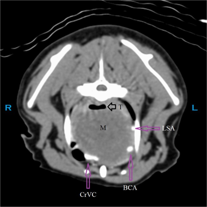
A 3.5-year-old castrated male domestic medium hair cat was evaluated for dry cough and labored breathing. A cranial mediastinal mass was seen on thoracic radiographs. On computed tomography, the mass displaced the cranial vena cava and dorsally displaced and compressed the intrathoracic trachea. The patient was taken to surgery for attempted mass removal. Intraoperatively, the mass was adhered to the cranial vena cava, aortic arch, left subclavian artery, and had partially engulfed the brachiocephalic trunk. The cat was euthanized and on necropsy the mass was found to invade the lumen of the vena cava and the tracheal lumen. An Aspergillus lentulus fungal granuloma was diagnosed histologically and by fungal culture and PCR. We highlight the difficulty in determining the extent of invasion with invasive aspergillosis and provide evidence that invasive aspergillosis can occur in otherwise healthy, young cats with no concurrent immunosuppressive treatments or comorbidities.
Keywords: Aspergillus Lentulus; cranial mediastinal mass; fungal granuloma.
© 2025 The Author(s). Journal of Veterinary Internal Medicine published by Wiley Periodicals LLC on behalf of American College of Veterinary Internal Medicine.
Conflict of interest statement
Authors declare no off‐label use of antimicrobials.
The authors declare no conflicts of interest.
Figures


Similar articles
-
Diffuse Osteosclerosis in a Young Cat Presenting With Chronic Nasal Congestion: A Case Report.Vet Med Sci. 2025 Sep;11(5):e70574. doi: 10.1002/vms3.70574. Vet Med Sci. 2025. PMID: 40833355 Free PMC article.
-
Pulmonary vein stenosis secondary to a mediastinal mass in a cat.Can Vet J. 2025 Apr;66(4):402-408. Can Vet J. 2025. PMID: 40170944 Free PMC article.
-
[A case of allergic bronchopulmonary mycosis caused by Triodiomyces crassus].Zhonghua Jie He He Hu Xi Za Zhi. 2025 May 12;48(5):464-469. doi: 10.3760/cma.j.cn112147-20240831-00523. Zhonghua Jie He He Hu Xi Za Zhi. 2025. PMID: 40300872 Chinese.
-
Morphological, functional and neurological outcomes of craniectomy versus cranial vault remodeling for isolated nonsyndromic synostosis of the sagittal suture: a systematic review.JBI Database System Rev Implement Rep. 2015 Sep;13(9):309-68. doi: 10.11124/jbisrir-2015-2470. JBI Database System Rev Implement Rep. 2015. PMID: 26470674
-
PET-CT for assessing mediastinal lymph node involvement in patients with suspected resectable non-small cell lung cancer.Cochrane Database Syst Rev. 2014 Nov 13;2014(11):CD009519. doi: 10.1002/14651858.CD009519.pub2. Cochrane Database Syst Rev. 2014. PMID: 25393718 Free PMC article.
References
-
- Davidson A. P., “Aspergillosis,” in Textbook of Veterinary Internal Medicine, vol. 1, 7th ed., ed. Ettinger S. J. and Feldman E. C. (Saunders Elsevier, 2010), 996–1002.
-
- Barrs V. R. and Dear J. D., “Aspergillosis and Penicilliosis,” in Greene's Infectious Diseases of the Dog and Cat, 5th ed., ed. Sykes J. E. (Elsevier, 2023), 1069–1093.
-
- Bueno D. J., Silva J. O., and Oliver G., “Mycoflora in Commercial Pet Foods,” Journal of Food Protection 64 (2001): 741–743. - PubMed
-
- Albhlal L. A., “Fungal Infection of the Nasal Cavity and Paranasal Sinuses: Review of 26 Cases,” Annals of Saudi Medicine 16 (1996): 615–621. - PubMed
Publication types
MeSH terms
LinkOut - more resources
Full Text Sources
Medical
Miscellaneous
