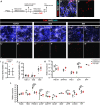
Anti-obesity compounds, Semaglutide and LiPR, and PrRP do not change the proportion of human and mouse POMC+ neurons
- PMID: 40802598
- PMCID: PMC12349008
- DOI: 10.1371/journal.pone.0329268
Anti-obesity compounds, Semaglutide and LiPR, and PrRP do not change the proportion of human and mouse POMC+ neurons
Abstract
Anti-obesity medications (AOMs) have become one of the most prescribed drugs in human medicine. While AOMs are known to impact adult neurogenesis in the hypothalamus, their effects on the functional maturation of hypothalamic neurons remain unexplored. Given that AOMs target neurons in the Medial Basal Hypothalamus (MBH), which play a crucial role in regulating energy homeostasis, we hypothesized that AOMs might influence the functional maturation of these neurons, potentially rewiring the MBH. To investigate this, we exposed hypothalamic neurons derived from human induced pluripotent stem cells (hiPSCs) to Semaglutide and lipidized prolactin-releasing peptide (LiPR), two anti-obesity compounds. Contrary to our expectations, treatment with Semaglutide or LiPR during neuronal maturation did not affect the proportion of anorexigenic, Pro-opiomelanocortin-expressing (POMC+) neurons. Additionally, LiPR did not alter the morphology of POMC+ neurons or the expression of selected genes critical for the metabolism or development of anorexigenic neurons. Furthermore, LiPR did not impact the proportion of adult-generated POMC+ neurons in the mouse MBH. Taken together, these results suggest that AOMs do not influence the functional maturation of anorexigenic hypothalamic neurons.
Copyright: © 2025 Jörgensen et al. This is an open access article distributed under the terms of the Creative Commons Attribution License, which permits unrestricted use, distribution, and reproduction in any medium, provided the original author and source are credited.
Conflict of interest statement
The authors have declared that no competing interests exist.
Figures


Similar articles
-
Hypothalamic POMC deficiency increases circulating adiponectin despite obesity.Mol Metab. 2020 May;35:100957. doi: 10.1016/j.molmet.2020.01.021. Epub 2020 Feb 7. Mol Metab. 2020. PMID: 32244188 Free PMC article.
-
Leptin signaling in POMC neurons regulates plasma leptin levels and is critical to mediate hypothalamic-pituitary-adrenal axis activation during fasting.Sci Rep. 2025 Jul 15;15(1):25574. doi: 10.1038/s41598-025-09990-0. Sci Rep. 2025. PMID: 40664708 Free PMC article.
-
The quantity, quality and findings of network meta-analyses evaluating the effectiveness of GLP-1 RAs for weight loss: a scoping review.Health Technol Assess. 2025 Jun 25:1-73. doi: 10.3310/SKHT8119. Online ahead of print. Health Technol Assess. 2025. PMID: 40580049 Free PMC article.
-
Prescription of Controlled Substances: Benefits and Risks.2025 Jul 6. In: StatPearls [Internet]. Treasure Island (FL): StatPearls Publishing; 2025 Jan–. 2025 Jul 6. In: StatPearls [Internet]. Treasure Island (FL): StatPearls Publishing; 2025 Jan–. PMID: 30726003 Free Books & Documents.
-
The Black Book of Psychotropic Dosing and Monitoring.Psychopharmacol Bull. 2024 Jul 8;54(3):8-59. Psychopharmacol Bull. 2024. PMID: 38993656 Free PMC article. Review.
References
MeSH terms
Substances
LinkOut - more resources
Full Text Sources
Miscellaneous

