. 2025 Aug 15;11(33):eadx1129.
doi: 10.1126/sciadv.adx1129. Epub 2025 Aug 13.

Structures of MmpL complexes reveal the assembly and mechanism of this family of transporters

Affiliations

Structures of MmpL complexes reveal the assembly and mechanism of this family of transporters

Zhemin Zhang et al. Sci Adv. .

Abstract

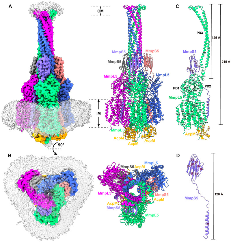
We coexpressed the mycobacterial membrane protein large 5 (MmpL5) transporter and MmpS5 adaptor proteins in Mycobacterium smegmatis and defined their structures from detergent-solubilized crude membranes. We observed that MmpL5 presents as a monomer in complex with the cytosolic meromycolate extension acyl carrier protein M (AcpM), where these AcpM-MmpL5 complexes generate regular two-dimensional arrays. We also provide structural information to show that M. smegmatis MmpL5 assembles as a trimer that interacts with MmpS5 and AcpM to form the tripartite complex AcpM-MmpL5-MmpS5 that spans both the inner and outer membranes of the mycobacterium. In addition, we found that MmpL5 and AcpM are able to form the trimeric AcpM-MmpL5 complex. The structural data reveal that the full-length MmpL5 trimer is capable of spanning the entire mycobacterial cell envelope to transport substrates. However, this assembly requires the presence of MmpS5 to stabilize secondary structural features of the MmpL5 periplasmic subdomains.


Figures

Fig. 1.
Fig. 1.. Duplicated AcpM-MmpL5 units form 2D arrays.
(A) Side view, (B) top view, and (C) bottom view of the cryo-EM maps and ribbon diagrams of duplicated AcpM-MmpL5 units within the 2D arrays. Individual MmpL5 molecules are in different colors. The periplasmic domain of MmpL5 consists of subdomains PD1, PD2, and PD3. Each AcpM molecule is colored orange. (D) Structure of an individual AcpM-MmpL5 unit within the AcpM-MmpL5 arrays. Each MmpL5 molecule forms an elongated channel (pink) that spans the pocket surrounded by TMs 7 to 10 at the outer leaflet of the cytoplasmic membrane and the cavity generated between subdomains PD1 and PD2 in the periplasm. In (A), (B), and (D), residues forming the PD3 subdomain of MmpL5 are missing in the structure. These residues are represented by dotted curves. (E) AcpM-MmpL5 interactions. The secondary structural elements of MmpL5 and AcpM are colored green and orange, respectively. Residues forming salt bridges at the interface between MmpL5 and AcpM are highlighted with sticks (as described in the “AcpM-MmpL5 interactions” section).
Fig. 2.
Fig. 2.. Structure of the tripartite AcpM-MmpL5-MmpS5 complex.
(A) Side view and (B) top view of the cryo-EM maps and ribbon diagrams of the AcpM-MmpL5-MmpS5 complex. The structure indicates that MmpL5 assembles as a trimer. The stoichiometry of AcpM:MmpL5:MmpS5 within the complex is in the form of a 3:3:3 molar ratio. Individual MmpL5 and MmpS5 molecules are colored differently. The AcpM molecules are colored orange and the detergent belts in the cryo-EM maps are colored gray. The inner and outer membrane regions are indicated as IM and OM, respectively. (C) Structure of a single unit of AcpM-MmpL5-MmpS5 within the trimeric complex. The structure indicates that the PD3 subdomain of MmpL5 forms an elongated hairpin of 125 Å in length, resulting in a vertical dimension of the MmpL5 protomer to be 215 Å. (D) Structure of a molecule of the MmpS5 adaptor. The structure indicates that MmpS5 is an elongated protein 120 Å long that is composed of an N-terminal TM domain of one single helix (TM) and a C-terminal periplasmic domain of eight β strands (β1 to β8).
Fig. 3.
Fig. 3.. Structure of the trimeric PD3 channel.
(A) Ribbon diagrams and electrostatic surface potentials of the side and top views of the trimer PD3 channel. The three PD3 domains of the MmpL5 trimer are in different colors. The blue (>10 kBT) and red (<−10 kBT) colors of the electrostatic surface potential indicate the positively and negatively charged areas of the protein, respectively, where kB is the Boltzmann constant and T is absolute temperature. White denotes area between 10 and −10 kBT. The needle-like apparatus of the PD3 channel is 125 Å long and 40 Å wide. (B) The interior of the central channel creates a pathway for transporting substrates. The channel, calculated by HOLE (http://holeprogram.org), is indicated by cyan dots. Residues forming the restriction sites and narrowest regions of the channel are represented with sticks. (C) The elongated substrate transport channel connecting the outer leaflet of the inner membrane up to the outer mycomembrane. The beginning of this channel is generated by a hydrophobic pocket created by TM7 to TM10 of MmpL5. This channel (colored blue) was calculated using the program CAVER (http://loschmidt.chemi.muni.cz/caver). In both (B) and (C), the restriction site is created by residues M615 and F619 of each PD3 subdomain of the MmpL5 trimer.
Fig. 4.
Fig. 4.. Interactions between MmpL5 and MmpS5.
Protomers 1, 2, and 3 of MmpL5 within the trimer are colored green, blue, and magenta, respectively. Molecules 1, 2, and 3 of MmpS5 are colored salmon, gray, and slate, respectively. The three AcpM molecules are colored orange. The TM helix of molecule 1 of MmpS5 (colored salmon) solely interacts with TM8 of protomer 1 of MmpL5 through hydrophobic interactions. The all–β strand periplasmic domain of molecule 1 of MmpS5 predominantly contacts protomer 2 of MmpL5 via hydrogen bonds and dipole-dipole interactions. This all–β strand domain of molecule 1 of MmpS5 also interacts with protomer 3 of MmpL5 via hydrogen bonds and charge-charge interactions. In addition, residues of the loop region of molecule 1 of MmpS5 contact protomers 1 and 2 of MmpL5 through hydrogen bonds, charge-charge, dipole-dipole, and hydrophobic interactions to further strengthen the binding. Residues participating in these interactions are in sticks. The hydrogen bonds are indicated with dotted lines.
Fig. 5.
Fig. 5.. Structure of the dipartite AcpM-MmpL5 complex.
(A) Side view and (B) top view of the cryo-EM maps and ribbon diagrams of the AcpM-MmpL5 complex. The structure indicates that MmpL5 assembles as a trimer. The stoichiometry of AcpM:MmpL5 within the complex is in the form of a 3:3 molar ratio. Individual MmpL5 molecules are colored differently. The AcpM molecules are colored orange, and the detergent belts in the cryo-EM maps are colored gray. The inner membrane region is indicated as IM. (C) Structure of a single unit of AcpM-MmpL5 within the trimeric complex. The structure indicates that the top half of the PD3 hairpin is unstructured.
Fig. 6.
Fig. 6.. Proposed mechanism for the assembly of the AcpM-MmpL5-MmpS5 trimeric complex.
In the absence of MmpS5, the AcpM-MmpL5 molecules form 2D arrays, which facilitate their lateral diffusion along the inner membrane of the mycobacterium. In the presence of MmpS5, MmpL5 forms a trimer and the tripartite AcpM-MmpL5-MmpS5 complex assembles. The trimer oligomerization of MmpL5 and the interaction between MmpL5 and MmpS5 stabilize the secondary structural elements of the three PD3 subdomains of the MmpL5 trimer, allowing them to create an elongated channel that protrudes toward the outer mycomembrane. The presence of the arabinogalactan (AG) and peptidoglycan (PG) layers create sufficient resistance to prevent the lateral movement of the trimeric AcpM-MmpL5-MmpS5 complex on the membrane plane. After the extrusion of substrates, the trimeric AcpM-MmpL5-MmpS5 complex can be dismantled by removing the three MmpS5 adaptor molecules to form the trimeric AcpM-MmpL5 complex. In this state, the secondary structural elements of subdomain PD3 of MmpL5 become unsteady and the top portion of the secondary structure of the hairpin becomes untraceable. The trimeric AcpM-MmpL5 complex then dismantles to form three monomeric AcpM-MmpL5 complexes, where the secondary structural elements of the entire PD3 subdomain of each MmpL5 molecule are untraceable and form a flexible loop. These AcpM-MmpL5 monomers then repack and form 2D arrays to stabilize the AcpM-MmpL5 complex structure.

Update of

Similar articles

References

    1. World Health Oganization, Global Tuberculosis Report 2024 (World Health Oganization, 2024).
    1. Wallis R. S., Maeurer M., Mwaba P., Chakaya J., Rustomjee R., Migliori G. B., Marais B., Schito M., Churchyard G., Swaminathan S., Hoelscher M., Zumla A., Tuberculosis—Advances in development of new drugs, treatment regimens, host-directed therapies, and biomarkers. Lancet Infect. Dis. 16, e34–e46 (2016). - PubMed
    1. World Health Oganization, Global tuberculosis report 2017 (World Health Oganization, 2017).
    1. Brennan P. J., Nikaido H., The envelope of mycobacteria. Annu. Rev. Biochem. 64, 29–63 (1995). - PubMed
    1. Cole S. T., Brosch R., Parkhill J., Garnier T., Churcher C., Harris D., Gordon S. V., Eiglmeier K., Gas S., Barry C. E. III, Tekaia F., Badcock K., Basham D., Brown D., Chillingworth T., Connor R., Davies R., Devlin K., Feltwell T., Gentles S., Hamlin N., Holroyd S., Hornsby T., Jagels K., Krogh A., McLean J., Moule S., Murphy L., Oliver K., Osborne J., Quail M. A., Rajandream M. A., Rogers J., Rutter S., Seeger K., Skelton J., Squares R., Squares S., Sulston J. E., Taylor K., Whitehead S., Barrell B. G., Deciphering the biology of Mycobacterium tuberculosis from the complete genome sequence. Nature 393, 537–544 (1998). - PubMed

MeSH terms

Substances

LinkOut - more resources