

Image Imputation with conditional generative adversarial networks captures clinically relevant imaging features on computed tomography
- PMID: 40802824
- PMCID: PMC12349720
- DOI: 10.1371/journal.pdig.0000970
Image Imputation with conditional generative adversarial networks captures clinically relevant imaging features on computed tomography
Abstract
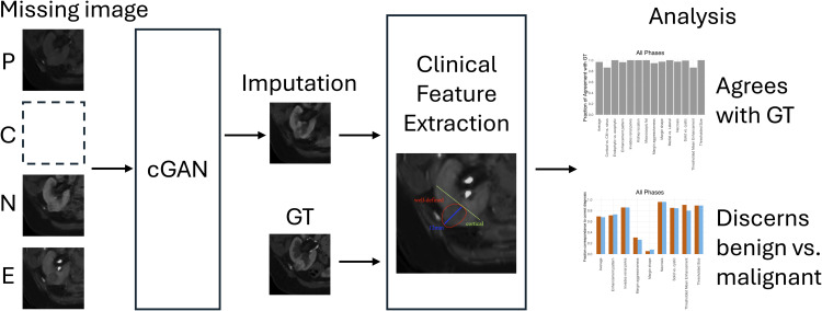
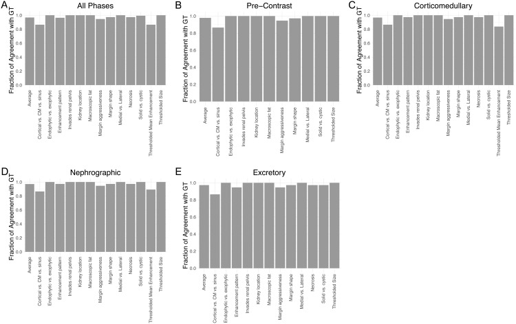
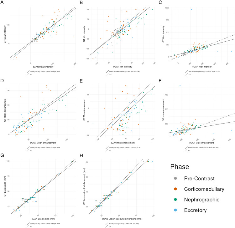
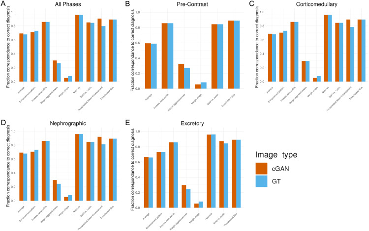
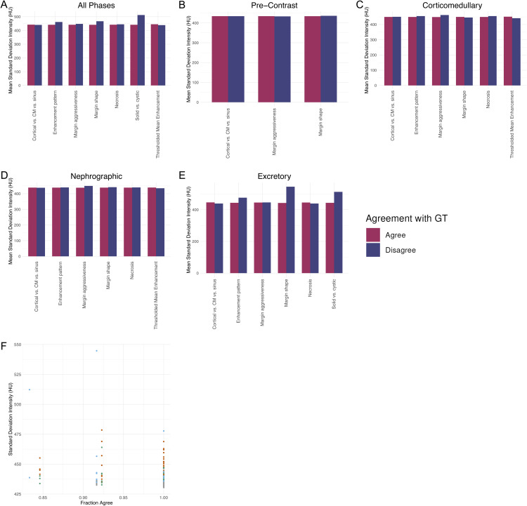
Kidney cancer is among the top 10 most common malignancies in adults, and is commonly evaluated with four-phase computed tomography (CT) imaging. However, the presence of missing or corrupted images remains a significant problem in medical imaging that impairs the detection, diagnosis, and treatment planning of kidney cancer. Deep learning approaches through conditional generative adversarial networks (cGANs) have recently shown technical promise in the task of imputing missing imaging data from these four-phase studies. In this study, we explored the clinical utility of these imputed images. We utilized a cGAN trained on 333 patients, with the task of the cGAN being to impute the image of any phase given the other three phases. We tested the clinical utility on the imputed images of the 37 patients in the test set by manually extracting 21 clinically relevant imaging features and comparing them to their ground truth counterpart. All 13 categorical clinical features had greater than 85% agreement rate between true images and their imputed counterparts. This high accuracy is maintained when stratifying across imaging phases. Imputed images also show good agreement with true images in select radiomic features including mean intensity and enhancement. Imputed images possess the features characteristic of benign or malignant diagnosis at an equivalent rate to true images. In conclusion, imputed images from cGANs have large potential for clinical use due to their ability to retain clinically relevant qualitative and quantitative features.
Copyright: © 2025 Rich et al. This is an open access article distributed under the terms of the Creative Commons Attribution License, which permits unrestricted use, distribution, and reproduction in any medium, provided the original author and source are credited.
Conflict of interest statement
I have read the journal's policy and the authors of this manuscript have the following competing interests: 1. Vinay Duddalwar is a consultant to Radmetrix, Roche, and Deeptek. The authors have no other relevant financial or non-financial interests to disclose.
Figures
Similar articles
-
Prescription of Controlled Substances: Benefits and Risks.2025 Jul 6. In: StatPearls [Internet]. Treasure Island (FL): StatPearls Publishing; 2025 Jan–. 2025 Jul 6. In: StatPearls [Internet]. Treasure Island (FL): StatPearls Publishing; 2025 Jan–. PMID: 30726003 Free Books & Documents.
-
Systemic pharmacological treatments for chronic plaque psoriasis: a network meta-analysis.Cochrane Database Syst Rev. 2017 Dec 22;12(12):CD011535. doi: 10.1002/14651858.CD011535.pub2. Cochrane Database Syst Rev. 2017. Update in: Cochrane Database Syst Rev. 2020 Jan 9;1:CD011535. doi: 10.1002/14651858.CD011535.pub3. PMID: 29271481 Free PMC article. Updated.
-
Comparison of Two Modern Survival Prediction Tools, SORG-MLA and METSSS, in Patients With Symptomatic Long-bone Metastases Who Underwent Local Treatment With Surgery Followed by Radiotherapy and With Radiotherapy Alone.Clin Orthop Relat Res. 2024 Dec 1;482(12):2193-2208. doi: 10.1097/CORR.0000000000003185. Epub 2024 Jul 23. Clin Orthop Relat Res. 2024. PMID: 39051924
-
Sexual Harassment and Prevention Training.2024 Mar 29. In: StatPearls [Internet]. Treasure Island (FL): StatPearls Publishing; 2025 Jan–. 2024 Mar 29. In: StatPearls [Internet]. Treasure Island (FL): StatPearls Publishing; 2025 Jan–. PMID: 36508513 Free Books & Documents.
-
Systemic pharmacological treatments for chronic plaque psoriasis: a network meta-analysis.Cochrane Database Syst Rev. 2021 Apr 19;4(4):CD011535. doi: 10.1002/14651858.CD011535.pub4. Cochrane Database Syst Rev. 2021. Update in: Cochrane Database Syst Rev. 2022 May 23;5:CD011535. doi: 10.1002/14651858.CD011535.pub5. PMID: 33871055 Free PMC article. Updated.
References
-
- Murphy A. CT renal mass (protocol) | Radiology Reference Article | Radiopaedia.org. n.d. [cited 2024 February 23]. https://radiopaedia.org/articles/ct-renal-mass-protocol-1?lang=us
-
- Galifer RB, Couture A, Dyon JF, Chappuis JP, Valla JS, Chavrier Y, et al. Solid tumors of the adrenal gland in children (excluding neuroblastomas). A study of a series of 18 cases. Chir Pediatr. 1989;30(5):209–14. - PubMed
-
- Pierorazio PM, Hyams ES, Tsai S, Feng Z, Trock BJ, Mullins JK, et al. Multiphasic enhancement patterns of small renal masses (≤4 cm) on preoperative computed tomography: utility for distinguishing subtypes of renal cell carcinoma, angiomyolipoma, and oncocytoma. Urology. 2013;81(6):1265–71. doi: 10.1016/j.urology.2012.12.049 - DOI - PMC - PubMed
LinkOut - more resources
Full Text Sources