. 2025 Aug 13;19(8):e0013401.
doi: 10.1371/journal.pntd.0013401. eCollection 2025 Aug.

Elucidating the functional role of the novel BdP50 protein and extracellular vesicles in the human erythrocyte infection by Babesia divergens

Affiliations

Elucidating the functional role of the novel BdP50 protein and extracellular vesicles in the human erythrocyte infection by Babesia divergens

Luis Miguel Gonzalez et al. PLoS Negl Trop Dis. .

Abstract

Babesia divergens is a blood-borne parasite that invades, replicates within and destroys red blood cells (RBCs) during its asexual life cycle, causing babesiosis in humans and cattle. This study focuses on BdP50, a putative B. divergens glycosylphosphatidylinositol-anchored protein involved in the parasite life cycle. BdP50 is found on the surface of B. divergens invasive parasites (merozoites) as well as on extracellular vesicles (Bd-derived EVs). These EVs are secreted by parasites cultured in fresh human RBCs and, in addition to BdP50, are enriched in human and parasite proteins, including proteins related to the parasite invasion process. BdP50 binds to RBCs and could mediate interactions of free merozoites and Bd-derived EVs with the host cell. Anti-BdP50 antibodies support this by blocking the BdP50 protein and inhibiting up to 88% of merozoite entry into naïve RBCs. This reinforces the role of BdP50 in parasite-host cell interactions and invasion. However, the inhibitory effect of BdP50 antibodies begins to gradually decrease slightly several hours after invasion, leading to a progressive increase in B. divergens infected RBCs over time. Consistent with these findings, our in vitro de novo infection assays showed that Bd-derived EVs, in addition to promoting parasite propagation, display proteins such as BdP50 that mimic the merozoite surface to likely attenuate the blocking effect of antibodies, thereby ensuring the parasite survival during subsequent rounds of invasion and growth. Given the role of BdP50 and Bd-derived EVs in the B. divergens life cycle, this study could have future implications for developing new approaches to interfere with parasite invasion proteins and Bd-derived EVs functions.


Conflict of interest statement

The authors have declared that no competing interests exist.

Figures

Fig 1
Fig 1. Identification of BdP50 in B. divergens cultures.
Western blot using B. divergens free merozoites (lanes 1 and 4), B. divergens iRBCs (lanes 2 and 5) and B. divergens supernatants (lanes: 3 and 6) from in vitro cultures, and serum against rBdP50Ct (lines 1, 2 and 3) or preimmune serum, as negative control (lanes 4, 5 and 6). One band of ⁓48 kDa (p48) and a dominant band of ⁓35 kDa (p35) (marked with arrows) were identified by anti-rBdP50Ct antibodies in B. divergens free merozoites, iRBCs and supernatants. Molecular mass markers are shown on the left.
Fig 2
Fig 2. BdP50 localizes at the apical end and around the surface of B. divergens parasites.
Immunofluorescent assays of intraerythrocytic and free parasites were performed by using anti-rBdP50Ct antibodies and preimmune rabbit serum as a negative control. Bound antibody was detected by using fluorescein isothiocyanate-conjugated anti–rabbit IgG antibodies. The preparations were counterstained with DAPI and examined by confocal microscopy. Samples were laser-stimulated at 488 and 405 nm. (A) Intraerythrocytic parasites were fixed with acetone-methanol (1:1) and probed with anti-rBdP50Ct antibodies. (B) Intraerythrocytic parasites were also probed with a preimmune rabbit serum. (C) Free merozoites were fixed with acetone-methanol (1:1) and probed with anti-rBdP50Ct antibodies. Free merozoites were also probed with preimmune rabbit sera. (E) Free extracellular merozoites were fixed with 2% paraformaldehyde and probed with anti-rBdP50Ct antibodies. Column 1 shows transmitted light images. Column 2 shows green fluorescent parasites. Column 3 shows the parasite DNA stained with DAPI. Column 4 shows all images overlaid.
Fig 3
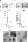
Fig 3. Characterization of B. divergens and human derived extracellular vesicles (EVs).
(A) Immunolocalization of BdP50 by Western blot using uRBC-derived EVs and Bd-derived EVs as targets, preimmune rabbit serum (lanes 1 and2) and anti-rBdP50Ct antibodies (lanes 3 and 4). Lane 1 and 3: uRBC-derived EVs. Lane 2 and 4: Bd-derived EVs. The BdP50 protein is predominantly found at 35 kDa (p35) and as a minor band at 48 kDa (p48) in Bd-derived EVs (black arrows). Molecular mass standards are shown on the left. The presence of BdP50 as a soluble protein in parasite culture supernatants free of EVs is ruled out by Western blot using anti-rBdP50Ct antibodies. Lane 5: recombinant His-tagged protein rBdP50 (~43 kDa) was used as a control. Lanes 6 and 7: 30 and 60 μg of concentrated B. divergens culture supernatants free of EVs, respectively. Babesia divergens merozoites (B) and Bd-derived EVs (C) were incubated either without (lanes 1 and 3) or with phosphatidylinositol-specific phospholipase C (PI-PLC) for 15 min (lanes 2 and 4). After centrifugation, pellets and supernatants were collected and analyzed by SDS-PAGE followed by immunoblotting with anti-BdP50 antibodies. Lanes 1 and 3: pellets and corresponding supernatants from untreated samples. Lanes 2 and 4: pellets and corresponding supernatants from PI-PLC-treated samples. Molecular mass markers are indicated on the left. (D) Nanoparticle tracking analysis (NTA) of EVs that were purified by ultracentrifugation and subsequent size exclusion chromatography from culture supernatants of B. divergens iRBCs and uRBCs. NTA showed Bd-derived EVs with a modal size between 123 ± 2.2 nm and 137.5 ± 2.6 nm, and uRBC-derived EVs with a modal size from 112.6 ± 3.8 to 116.7 ± 2.8 nm. Profile of size (in nm) concentration is shown. The highest concentration peak of Bd-derived EVs was between ~113 - ~ 139 nm, whereas for uRBC-derived EVs it was between ~113 - ~ 115 nm. (E) Negative staining and visualisation by transmission electron microscopy of Bd-derived EVs and uRBC-derived EVs. The size ranges of the purified EVs were consistent with the nanoparticle tracking analysis shown in panel A. Scale bars: 200 nm.
Fig 4
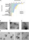
Fig 4. Most abundant B. divergens proteins in Bd-derived extracellular vesicles, including BdP50.
(A) The graph shows the 16 most abundant parasite proteins found in Bd-derived EVs per replicate. (*) B. divergens proteins involved in the red blood cell invasion process (+) Molecules that were also identified by the immunoscreening of the B. divergens cDNA library. Immunoelectron microscopy analysis of uRBC-derived EVs and Bd-derived EVs in glow-discharged collodion-carbon-coated copper grids fixed with 2% paraformaldehyde. (B) uRBC-derived EVs that were immunolabeled with preimmune rabbit serum. (C) Bd-derived EVs that were immunolabeled with a preimmune rabbit serum. (D) uRBC-derived EVs that were immunolabeled with anti-rBdP50Ct antibodies. (E) BdP50 is exclusively localized in Bd-derived EVs that were immunolabeled with anti-BdP50Ct antibodies. Bound antibodies were detected with a goat anti-rabbit conjugated to 15-nm gold particles. Scale bars: 200 and 100 nm.
Fig 5
Fig 5. Babesia divergens-derived EV treatment favours parasite invasion and growth in human RBCs.
Naïve RBC were pre-incubated for 18 h with Bd-derived EVs. Naïve RBCs were also incubated without exogenous Bd-derived EVs and used as a control. Babesia divergens free merozoites were then add to the pre-treated RBCs (with or without EVs) and parasitemia level was monitored using Giemsa-stained smears. Graphics A and B illustrate the significant differences in parasitemia between the group treated with Bd-derived EVs, which shows higher levels, and the group that did not receive any exogenous EVs. Each value represents the mean of the readings obtained from triplicates for each group. Error bars represent standard deviations of the mean levels of parasitemia, *p = 0.03, **p = 0.007.
Fig 6
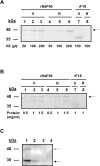
Fig 6. BdP50 is an erythrocyte-binding protein.
(A) Binding assay using increasing amounts of packed human RBCs, ranging from 50 to 200 µl, and a constant amount of the recombinant rBdP50 protein (0.5 mg/ml) (black arrow). (B) Binding assay using increasing amounts of the recombinant rBdP50 protein (black arrow), ranging from 0.5 to 1.5 mg/ml, and a constant amount of packed RBCs (100 µl). Recombinant rF18 protein is used as a negative control. Lanes 1, 2, and 3 show unbound (U) rBdP50 protein and lanes 4, 5 and 6 bound rBdP50 protein, as revealed by Western blot using anti-His-tag monoclonal antibodies. The recombinant protein rF18 does not bind to human erythrocytes (lanes 7 and 8). (C) A binding assay mixing B. divergens culture supernatants or B. divergens supernatants free of EVs with packet RBCs was analyzed by Western blot using anti-rBdP50 antibodies. Babesia divergens culture supernatant (lane 1). Total eluate from RBCs after a binding reaction of the B. divergens culture supernatant (lane 2). Specific bound proteins (lane 3). BdP50 protein is found in B. divergens culture supernatant and eluate samples, predominantly at 35 kDa (p35) (black arrow) and as a faint minor band at 48 kDa (p48) (open arrow). The specific bound protein in lane 3 also corresponds to the major product p35. Specific bound proteins were not observed in B. divergens culture supernatants free of EVs (lane 4). Molecular mass markers are shown on the left.
Fig 7
Fig 7. Purified anti-rBdP50 antibodies inhibit invasion of B. divergens merozoites in vitro.
Naïve human RBC were infected by B. divergens free merozoites and parasitemia monitored at 12 h, 24 h, 36 and after 48 h post invasion in the continued presence of purified IgG antibodies from the anti-rBdP50 serum (rBdP50 Ab) or IgG antibodies from the preimmune serum (PI Ab) at 2 mg ml-1. An in vitro wild-type control (WT), free of IgG antibodies, was also monitored. The percentage of parasitemia was monitored by examination of Giemsa-stained smears. A and B) After 12 h, anti BdP50 Ab reach their maximum invasion inhibition of 88.2%, which is significantly higher than those achieved with PI Ab or in the control (no antibodies). Each value represents the mean of the readings (% parasitemia) obtained from triplicate samples for each antibody and control (WT). Error bars represent standard deviations of the mean levels of parasitemia.

Similar articles

References

    1. Kumar A, O’Bryan J, Krause PJ. The Global Emergence of Human Babesiosis. Pathogens. 2021;10(11):1447. doi: 10.3390/pathogens10111447 - DOI - PMC - PubMed
    1. Hildebrandt A, Zintl A, Montero E, Hunfeld K-P, Gray J. Human Babesiosis in Europe. Pathogens. 2021;10(9):1165. doi: 10.3390/pathogens10091165 - DOI - PMC - PubMed
    1. Zintl A, Mulcahy G, Skerrett HE, Taylor SM, Gray JS. Babesia divergens, a bovine blood parasite of veterinary and zoonotic importance. Clin Microbiol Rev. 2003;16(4):622–36. doi: 10.1128/CMR.16.4.622-636.2003 - DOI - PMC - PubMed
    1. Gray JS, Ogden NH. Ticks, Human Babesiosis and Climate Change. Pathogens. 2021;10(11):1430. doi: 10.3390/pathogens10111430 - DOI - PMC - PubMed
    1. Conesa JJ, Sevilla E, Terrón MC, González LM, Gray J, Pérez-Berná AJ, et al. Four-Dimensional Characterization of the Babesia divergens Asexual Life Cycle, from the Trophozoite to the Multiparasite Stage. mSphere. 2020;5(5):e00928-20. doi: 10.1128/mSphere.00928-20 - DOI - PMC - PubMed

Substances

LinkOut - more resources