. 2025 Aug 13;21(8):e1011542.
doi: 10.1371/journal.pgen.1011542. eCollection 2025 Aug.

Polymerization and flanking domains of the bactofilin BacA collectively regulate stalk formation in Asticcacaulis biprosthecum

Affiliations

Polymerization and flanking domains of the bactofilin BacA collectively regulate stalk formation in Asticcacaulis biprosthecum

Maxime Jacq et al. PLoS Genet. .

Abstract

Bactofilins are a recently discovered class of cytoskeletal protein, widely implicated in subcellular organization and morphogenesis in bacteria and archaea. Several lines of evidence suggest that bactofilins polymerize into filaments using a central β-helical core domain, flanked by variable N- and C- terminal domains that may be important for scaffolding and other functions. In Asticcacaulis biprosthecum, the bactofilin BacA serves as a topological organizer of stalk synthesis, localizing to the stalk base and coordinating the synthesis of these long, thin extensions of the cell envelope. The easily distinguishable phenotypes of wild-type A. biprosthecum stalks and ΔbacA "pseudostalks" make this an ideal system for investigating how mutations in BacA affect its functions in morphogenesis. Here, we redefine the core domain of A. biprosthecum BacA using various bioinformatics and biochemical approaches to precisely delimit the N- and C- terminal domains. We then show that loss of these terminal domains leads to cells with severe morphological abnormalities, typically presenting a pseudostalk phenotype. BacA mutants lacking the N- and C- terminal domains also exhibit localization defects, implying that the terminal domains of BacA may be involved in its subcellular positioning, possibly through regulatory interactions with membrane-associated factors or other morphological proteins. We further show that point mutations that render BacA defective for polymerization lead to stalk synthesis defects. Overall, our study suggests that BacA's polymerization capacity and domain-mediated cellular positioning play a crucial role in the protein's function as a morphogenic regulator.


Conflict of interest statement

The authors have declared that no competing interests exist.

Figures

Fig 1
Fig 1. The bactofilin domain of A. biprosthecum BacA extends beyond the Pfam- predicted domain sequence, and forms filaments in vitro even in the absence of the BacA N- and C- terminal domains.
A) Multiple sequence alignment of bactofilin sequences from various species from the Caulobacteraceae family. The sequences are colored using a percentage-identity color scheme, which color-codes based on conservation (dark blue: above 80%; blue: above 60%; light blue: above 40%). The Pfam-predicted bactofilin domain (DUF583) is indicated with a magenta bracket, and the corrected bactofilin domain with a mauve bracket. B) Disorder plot of the A. biprosthecum BacA sequence, generated using SPOT-Disorder2, IUPred 3, Md2, and Espritz. Disorder scores were normalized from 0 to 1. Amino acid positions with a disorder score above 0.5 are considered disordered regions of the protein. A schematic of the BacA protein indicating its Pfam-predicted bactofilin domain (DUF583), is aligned with the disorder plot as reference. C) Left: Structural superposition of the structure of A. biprosthecum BacA predicted using AlphaFold (green), with its Pfam-delimited bactofilin domain (DUF583) in magenta. Right: Structural superposition of the predicted structure of A. biprosthecum BacA (green) with the NMR-resolved structure of C. crescentus BacA (PDB-ID: 2N3D, orange). D) High-resolution AFM images of purified BacA filaments and BacAΔNΔC. Scale bar = 1 µm. E) Bacterial two-hybrid (BACTH) assay in which pairs of proteins are fused to T18 and T25 fragments of adenylate cyclase and co-expressed in E. coli. Protein pairs that interact in situ enable the T18 and T25 fragments to carry out adenylate cyclase activity, which is reported as blue patches on beta-galactosidase culture plates. Here, T18-BacA constructs, with or without the BacA N- and C- terminal domains, are probed against full-length BacA fused to the T25 probe either on its N- or C-terminal. A strain co-expressing T18 and T25 fragments fused to interacting segments of a Leucine Zipper motif was used as a positive control (blue patch, bottom right). Negative controls (white patches) were performed against unfused T25 fragments.
Fig 2
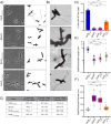
Fig 2. The disordered N- and C- terminal domains of BacA are required for normal morphogenesis of the stalk.
A) Phase contrast images (left) and corresponding cell silhouettes (right) of A. biprosthecum bacA-mVenus, ΔbacA, bacAΔN-mVenus and bacAΔC-mVenus, to analyze stalk morphology. Cells were grown in phosphate-limited (HIGG) medium (see Methods). Scale bars = 2 μm. B) Transmission electron microscopy of the same strains as in Panel A. Pseudostalks are indicated with red arrows. Scale bars = 2 μm. C) Summary statistics for data presented in Fig 2D-F. Data shown as mean ± SD. D) Percentage of cells with WT-like stalks. Based on phase images, cells were scored as having a WT-like stalk (i.e., a thin extension from the cell body). Cells exhibiting thick and aberrant pseudostalks were excluded (bacA-mVenus n = 3966; ΔbacA n = 2749; bacAΔN-mVenus n = 3034; bacAΔC-mVenus n = 3879). Data are presented as the mean percentage of cells with WT-like stalks. Error bars represent SD. (****p ≤ 0.001, ns = not significant p > 0.05; t-test). E) Distribution of stalk/pseudostalk lengths in the different strains (bacA-mVenus n = 575; ΔbacA n = 776; bacAΔN-mVenus n = 448; bacAΔC-mVenus n = 576). Data are represented as box and whisker plots and violin plots. (****p ≤ 0.001; t-test). F) Distribution of stalk/pseudostalk base diameter in the different strains based on atomic force microscopy images (bacA-mVenus n = 32; ΔbacA n = 13; bacAΔN-mVenus n = 15; bacAΔC-mVenus n = 23; see S3B Fig for representative images). Data are represented as box and whisker plots plots. (****p ≤ 0.001, ns = not significant p > 0.05; t-test).
Fig 3
Fig 3. The N- and C-terminal domains of BacA are necessary to localize BacA at the site of stalk synthesis but are not necessary for SpmX localization despite impaired interaction.
A) Merged phase contrast/fluorescence (left) and fluorescence (middle) microscopy images with localization heatmaps (right) of A. biprosthecum WT BacA-mVenus, BacAΔN-mVenus, and BacAΔC-mVenus. Red arrowheads indicate elongated foci of BacAΔC-mVenus. The number of cells analyzed is shown on the bottom left of each heatmap. Scale bars = 2 μm. B) Bacterial two-hybrid (BACTH) assays as presented in Fig 1E, with T18-BacA constructs with or without the BacA N- and C- terminal domains, probed against SpmX fused to the T25 probe either on its N- or C-terminal. C) Merged phase contrast/fluorescence (left) and fluorescence (middle) microscopy images with localization heatmaps (right) of SpmX-mCherry in A. biprosthecum WT, bacAΔN, and bacAΔC strains grown in 90 µM phosphate HIGG. The number of cells analyzed in each case is shown on the bottom left of each heatmap. Scale bars = 2 μm.
Fig 4
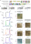
Fig 4. Various amino acid residues in the N-to-N and C-to-C interfaces are implicated in BacA polymerization.
A) Multiple sequence alignment of bactofilin sequences from A. biprosthecum, A. excentricus, C. crescentus, H. pylori, and T. thermophilus. The alignment is colored using the Clustal X color scheme, which color codes amino acid residues with shared properties. The red boxes indicate conserved amino acids that have been shown to be important for bactofilin polymerization in other species, selected for substitution in A. biprosthecum in this study. B) Structural representation of the BacA dimer derived from the BacA tetramer structure predicted using AlphaFold, showing either the N-to-N interface (left) or the C-to-C interface (right). Mutated residues are highlighted. C) Analytical size exclusion chromatography of purified BacA carrying various mutations. Elution profiles are color coded as follows - Black: WT BacA; Blue: BacAF134R; Green: BacAI56R; Orange: BacAL46R; Purple: BacAV79R. Single N-to-N interface mutants are displayed with dashed lines while double F134R and N-to-N interface mutants of BacA are displayed as solid lines. Subpanels [i] to [iv] highlight different constructs: [i] F134R; [ii] I56R, [iii] L46R, [iv] V79A. Curves are normalized such that the maximum intensity peak is 100. The expected elution ranges of monomers, dimers and multimers are represented above each curve. D) E. coli BACTH assay with T18-conjugated BacA constructs mutated in one (left) or both (right) polymerization interfaces, probed against the same constructs conjugated to T25, fusing each probe to either the N- or C-terminal of the BacA protein. Negative controls were performed against unfused T25 fragments. Positive controls (+) are shown on the bottom right of each plate.
Fig 5
Fig 5. The BacAF134R C-to-C interface polymerization mutant retains some functionality for stalk synthesis in vivo in A. biprosthecum.
A) Phase contrast, fluorescence and merged microscopy images of BacA-mVenus strains carrying various polymerization mutations, grown in phosphate-limited HIGG medium. For better visualization, LUTs were adjusted to reduce the brightness of the BacA-mVenus and BacA I56R-mVenus fluorescence signals, as their punctate localization showed much higher intensity compared to the diffuse signals in other strains. See S5B Fig for fluorescent images of these two strains with the same LUTs as the other mutants presented in this figure.Scale bars = 2 μm. B) Percentage of cells with different types of stalks for each of the BacA-mVenus polymerization mutant strains. Based on phase contrast images from three independent biological replicates, stalks were assessed and categorized either as WT-like stalks or pseudostalks. Data are presented as the mean percentage of cells with WT-like stalks or pseudostalks. Error bars represent SD. Total numbers of cells for each strain were as follows: WT n = 1940; F134R n = 1312; L46R n = 1817, I56R n = 1645; V79A n = 1483; L46R F134R n = 1308; I56R F134R n = 1907; V79A F134R n = 1746. C) Distribution of stalk/pseudostalk lengths in BacA-mVenus polymerization mutant strains. Error bars represent SD. Total numbers of cells for each strain were as follows: WT n = 189; F134R n = 63; L46R n = 192, I56R n = 113; V79A n = 167; L46R F134R n = 155; I56R F134R n = 38; V79A F134R n = 70).

Similar articles

References

    1. Wagstaff J, Löwe J. Prokaryotic cytoskeletons: protein filaments organizing small cells. Nat Rev Microbiol. 2018;16(4):187–201. doi: 10.1038/nrmicro.2017.153 - DOI - PubMed
    1. Amos LA, Löwe J. Overview of the Diverse Roles of Bacterial and Archaeal Cytoskeletons. Subcell Biochem. 2017;84:1–26. doi: 10.1007/978-3-319-53047-5_1 - DOI - PubMed
    1. Errington J. Bacterial morphogenesis and the enigmatic MreB helix. Nat Rev Microbiol. 2015;13(4):241–8. doi: 10.1038/nrmicro3398 - DOI - PubMed
    1. Margolin W. FtsZ and the division of prokaryotic cells and organelles. Nat Rev Mol Cell Biol. 2005;6(11):862–71. doi: 10.1038/nrm1745 - DOI - PMC - PubMed
    1. Ausmees N, Kuhn JR, Jacobs-Wagner C. The bacterial cytoskeleton: an intermediate filament-like function in cell shape. Cell. 2003;115(6):705–13. doi: 10.1016/s0092-8674(03)00935-8 - DOI - PubMed

LinkOut - more resources