

Phototherapeutic Keratectomy Combined with epikeratophakia for band keratopathy secondary to juvenile idiopathic arthritis-associated uveitis: A case report
- PMID: 40803253
- PMCID: PMC12361600
- DOI: 10.1016/j.ijscr.2025.111714
Phototherapeutic Keratectomy Combined with epikeratophakia for band keratopathy secondary to juvenile idiopathic arthritis-associated uveitis: A case report
Abstract
Introduction and importance: Band keratopathy (Band keratopathy, BK) is not a rare corneal disease. However, BK secondary to juvenile idiopathic arthritis-associated uveitis (Juvenile Idiopathic Arthritis-associated Uveitis, JIA-U) is not common. This case is to investigate the feasibility, safety, and efficacy of Phototherapeutic Keratectomy Combined (Phototherapeutic Keratectomy Combined, PTK) with Epikeratophakia (Epikeratophakia. EP) procedure for BK secondary to JIA-U.
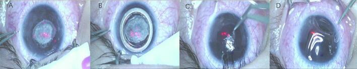
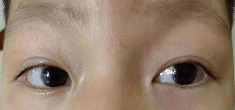
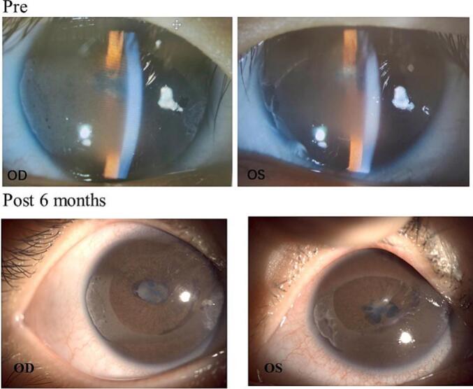
Case presentation: A 5-year-old boy was diagnosed with BK secondary to JIA-U. Due to the accumulation of calcareous ions in the central optical zone, the boy complained with blurred vision and photophobia. His two eyes were respectively treated with PTK combined with EP procedure using a transplanted lenticule obtained from small incision lenticule extraction (Small Incision Lenticule Extraction, SMILE) surgery. Uncorrected distance visual acuity (Uncorrected Distance Visual Acuity, UDVA), corrected distance visual acuity (Corrected Distance Visual Acuity, CDVA), and manifest refraction were assessed. Corneas were examined using a slit-lamp microscope, Pentacam corneal topography and anterior segment optical coherence tomography (AS-OCT).
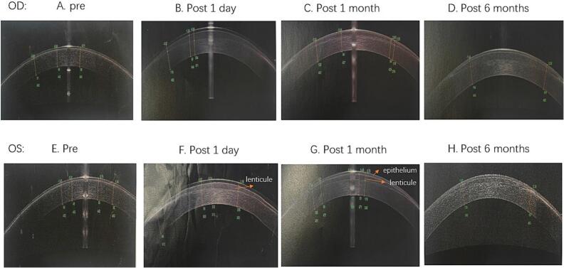
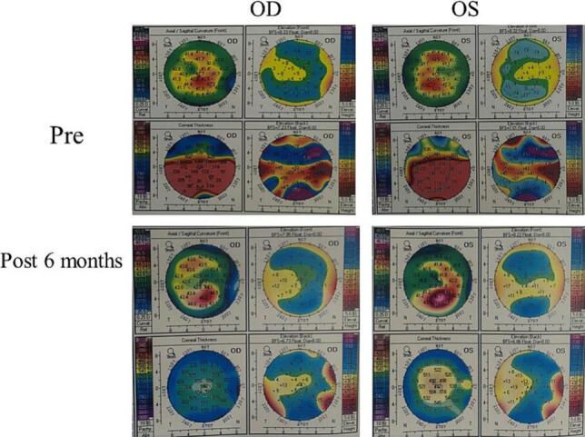
Clinical discussion: No complications and recurrence were observed during the follow-up period. The central corneal optical zone and transplanted lenticules kept transparent for all the follow-up period. AS-OCT showed that the corneal epithelial remodeling was completely achieved within 1 month, meanwhile, the transplanted lenticules kept transparent with a visible demarcation line and well-attached to the corneal stroma. Six months after surgery, central corneal thickness increased from pre-operative 503 μm to post-operative 619 μm in the right eye, and from pre-operative 503 μm to post-operative 549 μm in the left eye. The corneal topography showed perfect corneal shape and smooth surface. Three months and six months after surgery, the UDVA had been improved from 0.2 to 0.4 in the right eye and 0.25 to 0.4 in the left eye, the BCVA showed the same results as UDVA.
Conclusion: PTK combined with EP procedure is an innovative, feasible, safe and effective method for BK secondary to JIA-U, and therefore avoid the risk of hypermetropic shift of the refractive error and corneal dilation caused by repeated PTK procedure.
Keywords: Band keratopathy; Epikeratophakia technique; Femtosecond laser small incision lenticule extraction; Juvenile idiopathic arthritis-associated uveitis; Phototherapeutic Keratectomy.
Copyright © 2025 The Authors. Published by Elsevier Ltd.. All rights reserved.
Conflict of interest statement
Declaration of competing interest The author and co-authors have no conflicts of interest.
Figures
Similar articles
-
Photorefractive keratectomy with extended ablation zone for recurrent corneal erosion syndrome accompanied with refractive errors: a study of effectiveness, safety, and refractive outcomes.Front Med (Lausanne). 2025 Jul 18;12:1592539. doi: 10.3389/fmed.2025.1592539. eCollection 2025. Front Med (Lausanne). 2025. PMID: 40757202 Free PMC article.
-
Laser-assisted subepithelial keratectomy (LASEK) versus photorefractive keratectomy (PRK) for correction of myopia.Cochrane Database Syst Rev. 2016 Feb 22;2(2):CD009799. doi: 10.1002/14651858.CD009799.pub2. Cochrane Database Syst Rev. 2016. PMID: 26899152 Free PMC article.
-
Decreased vision due to scarring after phototherapeutic keratectomy.J Cataract Refract Surg. 2024 Oct 1;50(10):1094-1098. doi: 10.1097/j.jcrs.0000000000001529. J Cataract Refract Surg. 2024. PMID: 39313861
-
Laser-assisted subepithelial keratectomy (LASEK) versus laser-assisted in-situ keratomileusis (LASIK) for correcting myopia.Cochrane Database Syst Rev. 2017 Feb 15;2(2):CD011080. doi: 10.1002/14651858.CD011080.pub2. Cochrane Database Syst Rev. 2017. PMID: 28197998 Free PMC article.
-
Deep anterior lamellar keratoplasty versus penetrating keratoplasty for treating keratoconus.Cochrane Database Syst Rev. 2014 Jul 22;2014(7):CD009700. doi: 10.1002/14651858.CD009700.pub2. Cochrane Database Syst Rev. 2014. PMID: 25055058 Free PMC article.
References
-
- Spalton David J. A colour atlas of uveitis diagnosis. Br. J. Ophthalmol. 1985;69(8):636. doi: 10.1136/bjo.69.8.636. - DOI
Publication types
LinkOut - more resources
Full Text Sources
Research Materials
