. 2025 Oct 9;71(5):295-300.
doi: 10.1262/jrd.2025-025. Epub 2025 Aug 14.

A novel method of correlative light and electron microscopy in cryosectioning of bovine anterior pituitary tissue using NanoSuit CLEM

Affiliations

A novel method of correlative light and electron microscopy in cryosectioning of bovine anterior pituitary tissue using NanoSuit CLEM

Hiroya Kadokawa et al. J Reprod Dev. .

Abstract

Correlative light-electron microscopy (CLEM) combines fluorescence microscopy and scanning electron microscopy (SEM) to achieve nanoscale resolution while highlighting regions of interest identified by fluorescence microscopy. CLEM is becoming increasingly important in life sciences but traditionally requires highly dried samples to withstand the high vacuum of SEM. The NanoSuit method, which mimics native extracellular substances, was developed to address this limitation by encasing samples in a thin, vacuum-proof membrane, allowing SEM observation of live or wet multicellular organisms. While previous NanoSuit CLEM studies focused on formalin-fixed paraffin-embedded sections and cultured cells, cryosections had not yet been explored. In this study, NanoSuit CLEM with diluted NanoSuit solution was applied to cryosections of bovine anterior pituitary tissue. Secretory granules in gonadotrophs, which constitute less than 12% of anterior pituitary cells, were successfully visualized. However, other organelles remained unobserved due to fixation conditions. Therefore, NanoSuit CLEM enabled visualization of the ultrastructure of important cells in cryosections, even from large animals.

Keywords: Exocytosis; Gonadotroph; Luteinizing hormone; NanoSuit; Surface shield enhancer effect.


Conflict of interest statement

The authors have nothing to declare.

Figures
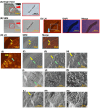
Fig. 1.
Fig. 1.
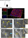
The NanoSuit method applied to cryosections for observation using scanning electron microscopy (SEM). The shape of blood vessels was used as a landmark near the region of interest and imaged with both a light microscope (A) and SEM (B). The red rectangles in images (A) and (B) indicate the regions shown at higher magnification in the corresponding panels. Immunohistochemistry of the tissue revealed LH, DAPI (as a nuclear counterstain), and their merged image (C). The blue rectangle in image (C) highlights the region shown at higher magnification in panel (D), where LH immunofluorescence, SEM, and a merged image are presented. Yellow and green arrows in (D) to (H) indicate LH-positive and LH-negative cells, respectively. The LH-positive and LH-negative cells were further observed by high-magnification SEM in images (I) to (N). Scale bars: 1 mm (low magnification in A, B); 400 μm (high magnification in A, B; and in C); 100 μm (D–F); 50 μm (G); 20 μm (H); 10 μm (I, L); 5 μm (J, M); and 2 μm (K, N).
Fig. 2.
Fig. 2.
(A) Significant differences in the diameter of secretory granules between LH-positive and LH-negative cells, as observed using the NanoSuit method and SEM. (B) Three-dimensional fluorescence immunohistochemistry images (220 μm in the X-axis, 180 μm in the Y-axis, and 10 μm in the Z-axis) showing LH (red), voltage-dependent anion channel 1 (VDAC1; green), and DNA counterstained with DAPI (blue). Note the absence of yellow fluorescence, indicating a lack of colocalization. (C, D) Yellow arrows and orange dotted lines indicate mitochondria occasionally detected by SEM in macerated sections of the same tissues coated with NanoSuit solution. The blue rectangle in image (C) indicates the region shown at higher magnification in image (D). In these mitochondria, only the outer membrane was visible, as shown in the upper right orange panel. The inner membrane and other organelles, such as the Golgi apparatus and endoplasmic reticulum, were not observed. (E, F) Yellow arrows and orange dotted lines indicate mitochondria occasionally detected by SEM in non-macerated sections of the same tissues coated with NanoSuit solution. The blue rectangle in image (E) indicates the region shown at higher magnification in image (F). As in the macerated sections, only the outer membrane was visible, as shown in the upper right orange panel (the pink arrow indicates the signal of a gold-labeled antibody), while the inner membrane and other organelles—such as the Golgi apparatus and endoplasmic reticulum—were not observed. Scale bars: 20 μm (C), 10 μm (E), 5 μm (D), and 2 μm (F).

References

    1. Timmermans FJ, Otto C. Contributed review: Review of integrated correlative light and electron microscopy. Rev Sci Instrum 2015; 86: 011501. - PubMed
    1. Takaku Y, Suzuki H, Ohta I, Tsutsui T, Matsumoto H, Shimomura M, Hariyama T. A ‘NanoSuit’ surface shield successfully protects organisms in high vacuum: observations on living organisms in an FE-SEM. Proc Biol Sci 2015; 282: 282. - PMC - PubMed
    1. Kawasaki H, Itoh T, Takaku Y, Suzuki H, Kosugi I, Meguro S, Iwashita T, Hariyama T. The NanoSuit method: a novel histological approach for examining paraffin sections in a nondestructive manner by correlative light and electron microscopy. Lab Invest 2020; 100: 161–173. - PMC - PubMed
    1. Itoh T, Yamada S, Ohta I, Meguro S, Kosugi I, Iwashita T, Itoh H, Kanayama N, Okudela K, Sugimura H, Misawa K, Hariyama T, Kawasaki H. Identifying active progeny virus particles in formalin-fixed, paraffin-embedded sections using correlative light and scanning electron microscopy. Lab Invest 2023; 103: 100020. - PubMed
    1. Sakano Y, Kawasaki H. Visualization of molluscum contagiosum virus in FFPE skin sections using NanoSuit-CLEM: Ultrastructural evidence of viral spread via skin barrier disruption. Immun Inflamm Dis 2025; 13: e70212. - PMC - PubMed

MeSH terms