

miR-221 activates Sox11 to reduce brain injury after intracerebral hemorrhage via inhibiting neuroinflammation
- PMID: 40804093
- PMCID: PMC12350757
- DOI: 10.1038/s41598-025-15239-7
miR-221 activates Sox11 to reduce brain injury after intracerebral hemorrhage via inhibiting neuroinflammation
Abstract
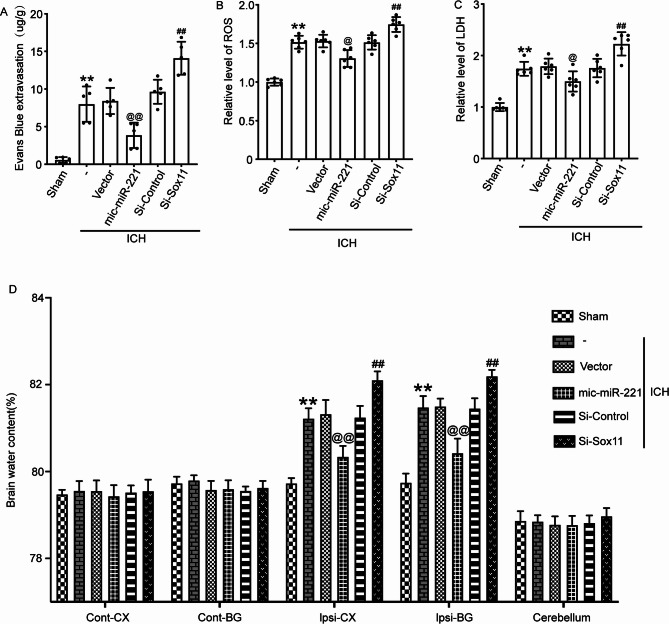
As a global public health issue, intracerebral hemorrhage (ICH) is characterized by high morbidity and mortality. Brain injury following ICH is composed of primary and secondary injury, with the latter being more severe and resulting in increased apoptosis. Sox11 (sex-determining region Y-related high-mobility-group 11), a vital member of the Sox gene family, is broadly discovered in the developing nervous system and may have a vital impact on neurogenesis, neuronal survival, and neurite outgrowth. The level and impacts of Sox11 in brain with ICH remain indistinct. The major objective of the current work was to explore the spatiotemporal expression of Sox11 and its roles in secondary brain injury under the ICH impairment. The ICH rat model was established by injecting autologous blood into the right basal ganglia of male Sprague-Dawley rats. It was observed that Sox11 expression was notably elevated in brain tissue after ICH. The enhancement of Sox11 expression through miR-221 reduced neuronal apoptosis and inflammation in the affected rats. Furthermore, overexpression of Sox11 mitigated ICH-induced brain edema, blood-brain barrier disruption, and cognitive impairments. In contrast, Sox11 knockdown resulted in opposing effects. These findings highlight the crucial role of Sox11 in alleviating secondary brain injury following ICH. Thus, upregulating Sox11 presents a promising therapeutic strategy to reduce secondary brain injury in clinical ICH cases.
Keywords: Intracerebral hemorrhage; MiR-221; Neuroinflammation; Secondary brain injury; Sox11.
© 2025. The Author(s).
Conflict of interest statement
Declarations. Ethics approval and consent to participate: All the experiments were authorized by the Institutional Animal Care Committee of Zhejiang Provincial people’s hospital and were conducted based on the guidelines of the National Institutes of Health on the care and use of animals. The study is reported in accordance with ARRIVE guidelines. Competing interests: The authors declare no competing interests.
Figures


Similar articles
-
miR-210 Regulates Autophagy Through the AMPK/mTOR Signaling Pathway, Reduces Neuronal Cell Death and Inflammatory Responses, and Enhances Functional Recovery Following Cerebral Hemorrhage in Mice.Neurochem Res. 2025 Jun 5;50(3):180. doi: 10.1007/s11064-025-04434-7. Neurochem Res. 2025. PMID: 40471451 Free PMC article.
-
Microglial STING activation promotes neuroinflammation and pathological changes in experimental mice with intracerebral haemorrhage.Acta Pharmacol Sin. 2025 Sep;46(9):2376-2392. doi: 10.1038/s41401-025-01540-8. Epub 2025 Apr 8. Acta Pharmacol Sin. 2025. PMID: 40200123
-
Transient receptor potential vanilloid 1 inhibition reduces brain damage by suppressing neuronal apoptosis after intracerebral hemorrhage.Brain Pathol. 2024 Sep;34(5):e13244. doi: 10.1111/bpa.13244. Epub 2024 Feb 2. Brain Pathol. 2024. PMID: 38308041 Free PMC article.
-
MicroRNAs as biomarkers in spontaneous intracerebral hemorrhage: A systematic review of recent clinical evidence.Clin Neurol Neurosurg. 2022 Feb;213:107130. doi: 10.1016/j.clineuro.2022.107130. Epub 2022 Jan 14. Clin Neurol Neurosurg. 2022. PMID: 35066247
-
Revisiting the role of the complement system in intracerebral hemorrhage and therapeutic prospects.Int Immunopharmacol. 2023 Oct;123:110744. doi: 10.1016/j.intimp.2023.110744. Epub 2023 Aug 7. Int Immunopharmacol. 2023. PMID: 37552908
References
-
- Zhang, S. et al. AAV/BBB-Mediated gene transfer of CHIP attenuates brain injury following experimental intracerebral hemorrhage. Transl. Stroke Res.11 (2), 296–309 (2020). - PubMed
-
- Chu, H. et al. Lactate dehydrogenase predicts early hematoma expansion and poor outcomes in intracerebral hemorrhage patients. Transl. Stroke Res.10 (6), 620–629 (2019). - PubMed
-
- van Asch, C. J. et al. Incidence, case fatality, and functional outcome of intracerebral haemorrhage over time, according to age, sex, and ethnic origin: a systematic review and meta-analysis. Lancet Neurol.9 (2), 167–176 (2010). - PubMed
MeSH terms
Substances
LinkOut - more resources
Full Text Sources
Research Materials
Miscellaneous
