. 2025 Aug 13;15(1):29716.
doi: 10.1038/s41598-025-14123-8.

Methylation-induced suppression of BEX1 activates AKT/ERK/STAT3 signaling pathways regulating cell cycle and apoptosis in glioma

Affiliations

Methylation-induced suppression of BEX1 activates AKT/ERK/STAT3 signaling pathways regulating cell cycle and apoptosis in glioma

Li-Zhi Xiao et al. Sci Rep. .

Abstract

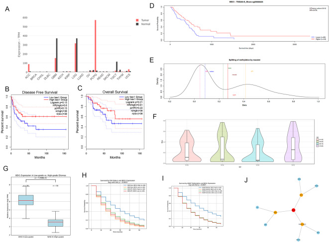
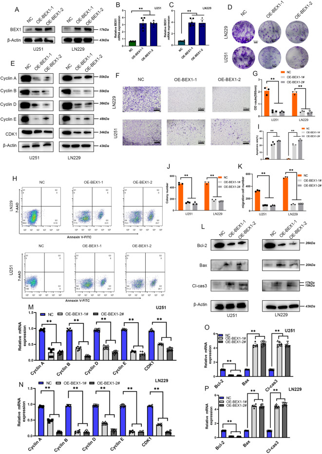
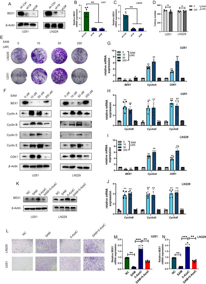
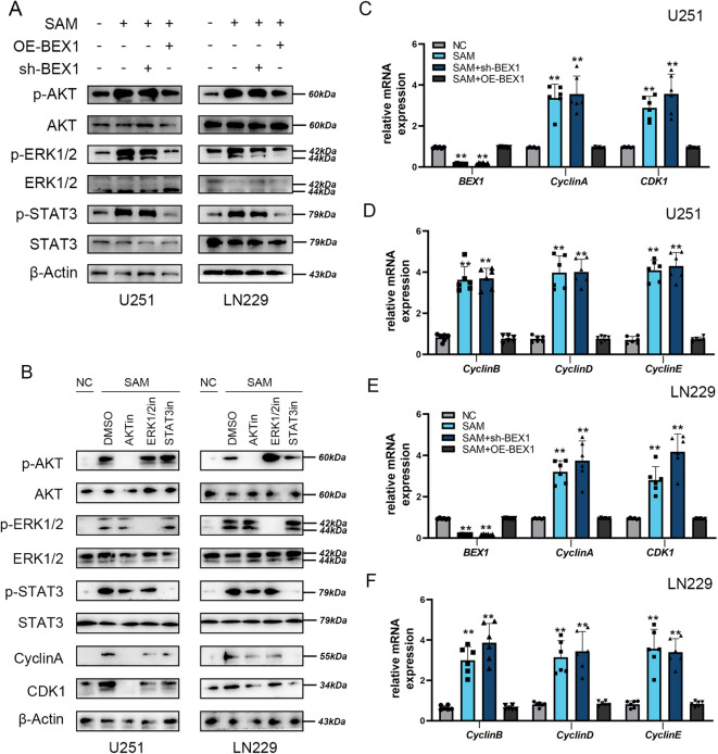
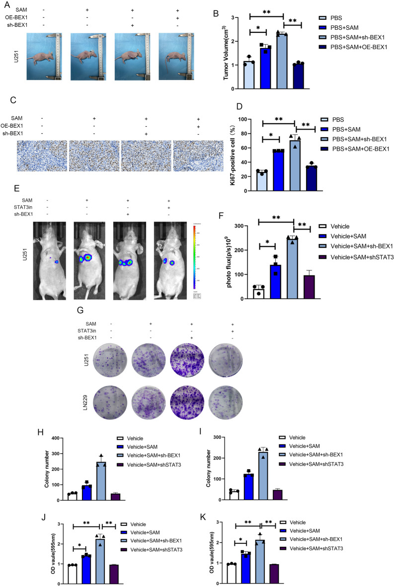
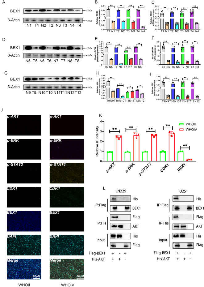
Gliomas are highly aggressive brain tumors with complex molecular characteristics. The role of BEX1 methylation in gliomas is not well understood, despite its potential implications for tumor biology and therapy. Investigating this relationship could uncover critical mechanisms underlying glioma pathogenesis and highlight therapeutic targets. This study aims to elucidate the specific mechanisms by which BEX1 methylation regulates the cell cycle and apoptosis in glioma. We conducted bioinformatics analyses to assess BEX1 expression differences in glioma using tissue samples, followed by validation through Western blot and qRT-PCR. Functional assays in glioma cell lines were performed, employing gene transfection and small molecule inhibitors to further explore BEX1's role in the AKT/ERK/STAT3 signaling pathways. Our findings reveal that BEX1 is significantly downregulated in gliomas due to promoter methylation, which in turn activates the AKT/ERK/STAT3 signaling cascade, leading to alterations in cell cycle regulation and apoptosis. Targeting BEX1 methylation presents a promising therapeutic avenue for glioma treatment. This study provides valuable insights into the mechanisms of BEX1 in glioma, paving the way for clinical translation and further research.

Keywords: BEX1; Clinical translation; Giloma; Methylation; PI3K/ERK/STAT3.


Conflict of interest statement

Declarations. Competing interests: The authors declare no competing interests.

Figures

Fig. 1
Fig. 1
Bioinformatics Analysis of BEX1 Expression in Glioma Patients. (A) Pan-cancer analysis showing significant downregulation of BEX1 in high-grade and low-grade gliomas. (B, C) Survival analyses indicating poorer disease-free survival and overall survival in cohorts with low BEX1 expression compared to those with high BEX1 expression. (D) Methylation analysis revealing worse survival outcomes in patients with low methylation levels. (E) Binary classification of patients based on critical methylation thresholds presented visually. (F) Violin plots illustrating the distribution of CpG methylation levels in relation to clinical factors such as age and sex. (G) BEX1 mRNA expression levels were analyzed in a cohort of glioma patients (n = 50), categorized by histopathological grade. A significant downregulation of BEX1 was observed in WHO IV gliomas compared to WHO II (p < 0.0001, unpaired two-tailed t-test). Data are presented as mean ± SEM. (H) Patients with IDH-mutant gliomas were divided into high and low BEX1 expression groups based on the median expression level. Higher BEX1 expression was associated with improved overall survival (p < 0.01, Log-rank test). (I) In MGMT-methylated gliomas, patients with high BEX1 expression also exhibited significantly longer survival than those with low BEX1 expression (p < 0.05). The number of patients in each group is indicated. (J) The network illustrates the potential interactions between BEX1 and associated signaling proteins, including AKT1, MAPK1 (ERK2), and STAT3. Nodes represent proteins and edges represent evidence-based predicted associations. Interaction confidence scores were set to medium (≥ 0.4). The network was visualized using STRING v12.
Fig. 2
Fig. 2
BEX1 overexpression inhibits proliferation, migration, and cell cycle progression while promoting apoptosis in glioma cells. (A–C) Western blot and qRT-PCR confirmation of BEX1 overexpression in U251 and LN229 cells using two constructs (OE-BEX1-1 and OE-BEX1-2). (D) Colony formation assay showing reduced colony numbers upon BEX1 overexpression. (E) Western blot analysis of Cyclin A, B, D, E, and CDK1 expression. (F) Transwell migration assay showing reduced migration in BEX1-overexpressing cells. (G) CCK-8 assay indicating reduced proliferation after BEX1 overexpression. (H–I) Representative flow cytometry dot plots and corresponding quantification of apoptosis in U251 and LN229 cells following BEX1 overexpression. Apoptotic status was assessed using Annexin V-FITC and 7-AAD staining. The quadrants represent: Q1 (Annexin V⁻/7-AAD⁺): necrotic cells. Q2 (Annexin V⁺/7-AAD⁺): late apoptotic cells. Q3 (Annexin V⁻/7-AAD⁻): viable/live cells. Q4 (Annexin V⁺/7-AAD⁻): early apoptotic cells. The proportion of early and late apoptotic cells (Q2 + Q4) was significantly increased in BEX1-overexpressing groups compared to the control. Data are presented as mean ± SEM (n = 3); **P < 0.01, *P < 0.05 (one-way ANOVA). (J) Quantification of colony formation data. (K) Quantification of transwell migration data. (L) Western blot analysis of apoptosis-related proteins: Bcl-2, Bax, and cleaved caspase-3 (Cl-cas3). (M–N) qRT-PCR analysis of cell cycle genes (Cyclins A–E and CDK1) in U251 and LN229 cells. (O–P) qRT-PCR analysis of apoptosis-related genes (Bcl-2, Bax, Cl-cas3) in U251 and LN229 cells. Data are presented as mean ± SEM; *P < 0.05, **P < 0.01.
Fig. 3
Fig. 3
Methylation suppresses BEX1 expression and promotes proliferation and migration in glioma cells. (A–C) Western blot and qRT-PCR showing BEX1 knockdown efficiency using two shRNAs in U251 and LN229 cells. (D) Colony formation under 0 or 200 µM SAM treatment shows no significant difference in colony number. (E) Crystal violet staining of colonies after SAM treatment (0–200 µM), showing increased colony formation at 50 µM. (F) Western blot showing downregulation of BEX1 and upregulation of Cyclins A–E and CDK1 with increasing SAM doses. (G–J) qRT-PCR results validating BEX1 suppression and upregulation of cell cycle genes (Cyclin A–E, CDK1) under SAM treatment in U251 (G–H) and LN229 (I–J) cells. (K) Western blot showing BEX1 expression in cells treated with SAM, 5-AzaC, or their combination. (L) Transwell assays showing that SAM enhances migration, while 5-AzaC suppresses it; combination treatment partially reverses SAM effects. (M–N) qRT-PCR analysis confirming BEX1 expression patterns in U251 (M) and LN229 (N) cells under the four treatment conditions (NC, SAM, 5-AzaC, SAM + 5-AzaC). Data are presented as mean ± SEM. *P < 0.05, **P < 0.01, ***P < 0.001 (one-way ANOVA with Tukey’s post hoc test).
Fig. 4
Fig. 4
Methylation suppresses BEX1 expression and activates the AKT/ERK/STAT3 signaling pathway to regulate the cell cycle in glioma cells. (A) Western blot analysis of p-AKT, p-ERK1/2, and p-STAT3 in U251 and LN229 cells following SAM-induced methylation, with or without BEX1 knockdown or overexpression. Total AKT, ERK1/2, and STAT3 served as internal references. (B) Inhibitor validation assays: U251 and LN229 cells were treated with SAM and specific inhibitors of AKT (AKTi), ERK1/2 (ERK1/2i), or STAT3 (STAT3in). The expression levels of phosphorylated proteins and downstream regulators (Cyclin A, CDK1) were detected by Western blot. (C–D) qRT-PCR quantification of BEX1, CyclinA, CDK1 (C), and CyclinB, CyclinD, CyclinE (D) expression in U251 cells across four groups (NC, SAM, SAM + shBEX1, SAM + OE-BEX1). (E–F) Corresponding mRNA expression profiles in LN229 cells. Data are presented as mean ± SEM (n = 3 per group). *P < 0.01, P < 0.05 compared with NC or as indicated (one-way ANOVA with Tukey’s post hoc test).
Fig. 5
Fig. 5
Methylation and BEX1 Influence Glioma Metastasis in Animal Models. (A) Subcutaneous tumor formation experiments showing increased tumor burden with methylation and BEX1 knockdown, contrasted with reduced burden upon BEX1 overexpression. (B) Relative quantification of tumor burdens. (C–D) Ki67 immunohistochemical staining and quantification of proliferating cells in xenograft tissues across indicated groups. (E) In vivo imaging of pulmonary metastasis revealing enhanced metastatic capability associated with methylation and BEX1 knockdown, mitigated by STAT3 pathway inhibition. (F) Relative quantification of lung metastasis outcomes. (G) Representative colony formation assay in U251 and LN229 cells under different conditions (Vehicle, SAM, SAM + sh-BEX1, SAM + sh-STAT3). (H–I) Quantification of colony numbers in U251 (H) and LN229 (I) cells. (J–K) CCK-8 assay showing cell proliferation (OD 450 nm) of U251 (J) and LN229 (K) cells under the same treatments. Data are presented as mean ± SEM. *P < 0.05, **P < 0.01 (one-way ANOVA with Tukey’s post hoc test).
Fig. 6
Fig. 6
Expression Analysis of BEX1 in Glioma Tissue Samples. (A-F) Western blot analysis confirming significantly reduced BEX1 protein levels in tumor tissues compared to adjacent non-tumor tissues from 12 glioma patients. (G-I) qRT-PCR analysis validating significantly decreased BEX1 mRNA levels in tumor tissues relative to adjacent non-tumor samples across all cases. (J) Representative immunofluorescence images of WHO grade II and IV glioma tissues stained for p-AKT, p-ERK, p-STAT3, CDK1, BEX1, and DAPI. (K) Quantification of relative fluorescence intensity showing increased p-AKT, p-ERK, p-STAT3, and CDK1, and decreased BEX1 in WHO grade IV gliomas (n = 3). (L) Co-immunoprecipitation in LN229 and U251 cells co-transfected with Flag-BEX1 and His-AKT. BEX1 physically associates with AKT, as shown by reciprocal IP with anti-Flag and anti-His antibodies. Data are shown as mean ± SEM. *P < 0.05, **P < 0.01; ns: not significant.

Similar articles

References

    1. Han, L. et al. SLC1A5 enhances malignant phenotypes through modulating ferroptosis status and immune microenvironment in glioma. Cell. Death Dis.13, 1071. 10.1038/s41419-022-05526-w (2022). - PMC - PubMed
    1. Lim, M., Xia, Y., Bettegowda, C. & Weller, M. Current state of immunotherapy for glioblastoma. Nat. Rev. Clin. Oncol.15, 422–442. 10.1038/s41571-018-0003-5 (2018). - PubMed
    1. Stupp, R. et al. Effect of Tumor-Treating fields plus maintenance Temozolomide vs maintenance Temozolomide alone on survival in patients with glioblastoma: A randomized clinical trial. JAMA318, 2306–2316. 10.1001/jama.2017.18718 (2017). - PMC - PubMed
    1. Chen, Z. et al. Roles of TRPM channels in glioma. Cancer Biol. Ther.25, 2338955. 10.1080/15384047.2024.2338955 (2024). - PMC - PubMed
    1. Wei, S. et al. Antitumor activity of a Mitochondrial-Targeted HSP90 inhibitor in gliomas. Clin. Cancer Res.28, 2180–2195. 10.1158/1078-0432.CCR-21-0833 (2022). - PMC - PubMed

LinkOut - more resources