. 2025 Aug 13;8(1):245.
doi: 10.1038/s42004-025-01644-6.

In vivo crosslinking and effective 2D enrichment for proteome wide interactome studies

Affiliations

In vivo crosslinking and effective 2D enrichment for proteome wide interactome studies

Philipp Bräuer et al. Commun Chem. .

Abstract

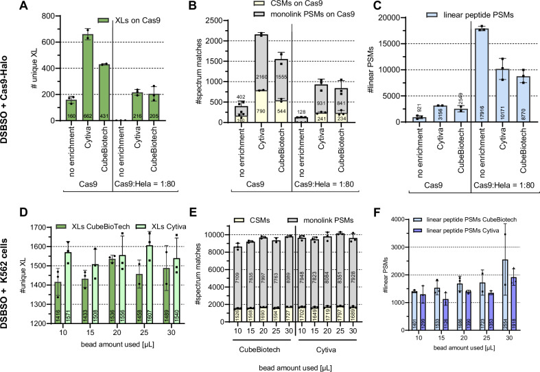
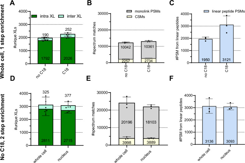
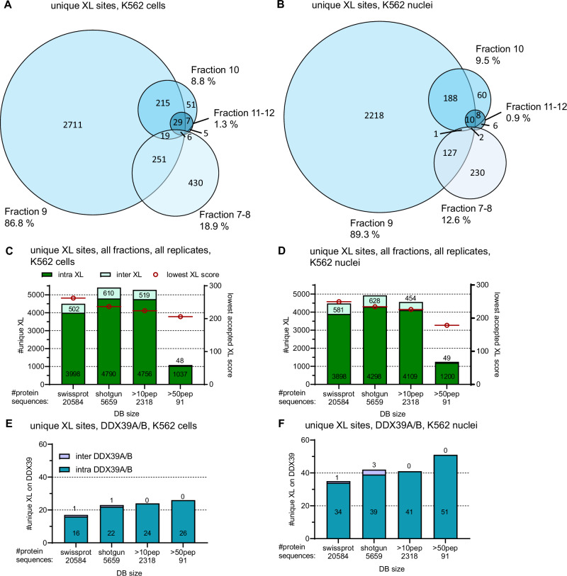
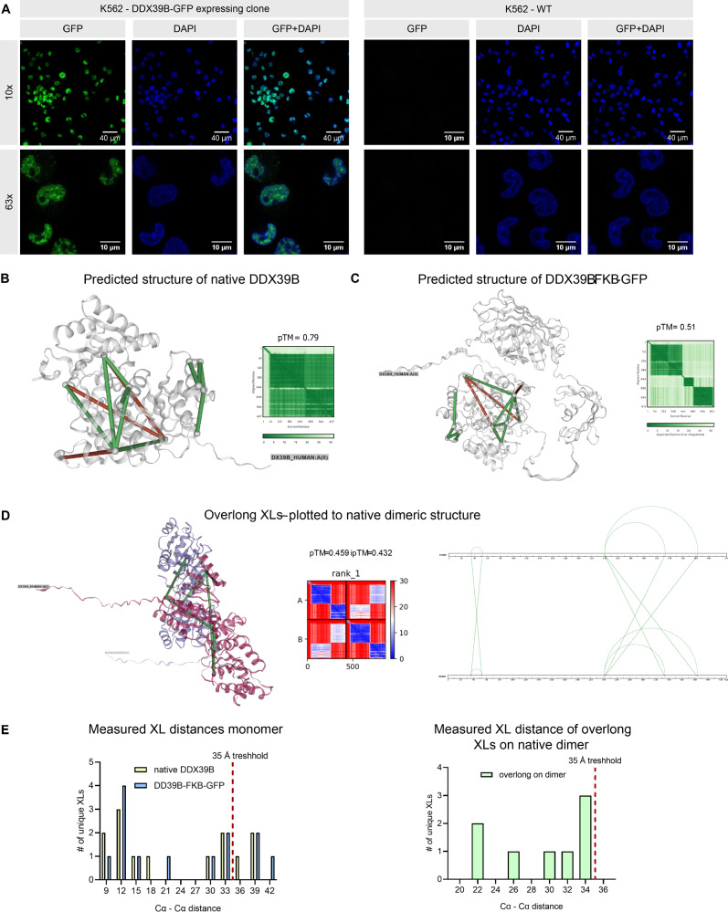
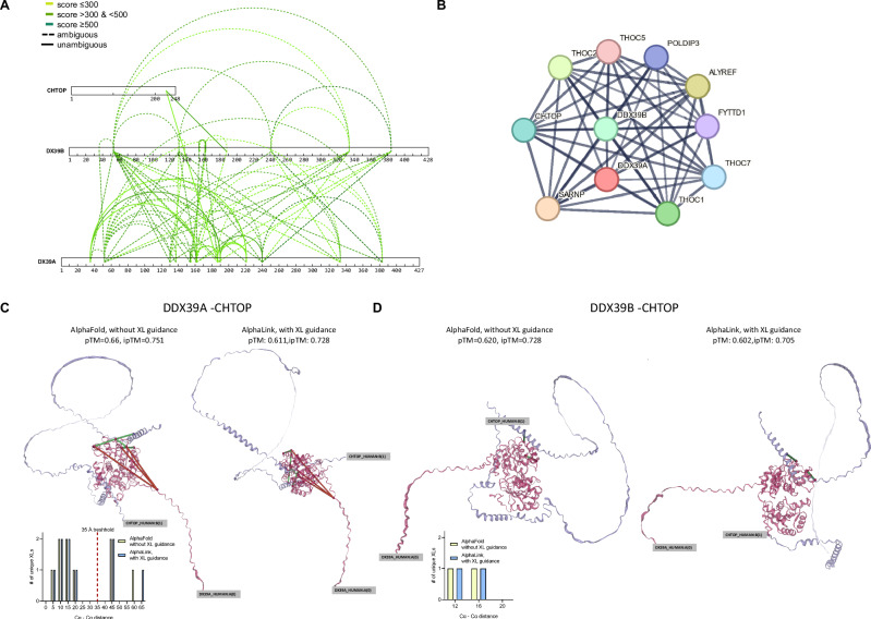
Cross-linking mass spectrometry has evolved as a powerful technique to study protein-protein interactions and to provide structural information. Low reaction efficiencies, and complex matrices lead to challenging system wide crosslink analysis. We improved and streamlined an Azide-A-DSBSO based in vivo crosslinking workflow employing two orthogonal effective enrichment steps: Affinity enrichment and size exclusion chromatography (SEC). Combined, they allow an effective enrichment of DSBSO containing peptides and remove the background of linear as well as mono-linked peptides. We found that the analysis of a single SEC fraction is effective to yield ~90% of all crosslinks, which is important whenever measurement time is limited, and sample throughput is crucial. Our workflow resulted in more than 5000 crosslinks from K562 cells and generated a comprehensive PPI network. From 393 PPI found within the nucleus, 56 are novel. We further show, that by applying DSBSO to nuclear extracts we yield more crosslinks on lower abundant proteins and showcase this on the DEAD-box RNA helicase DDX39B which is predominantly expressed in the nucleus. Our data indicates that DDX39B might be present in monomeric and dimeric forms together with DDX39A within the nuclear extracts analyzed.


Conflict of interest statement

Competing interests: The authors declare no competing interests.

Figures

Fig. 1
Fig. 1. Graphical workflow representation.
Cells or isolated nuclei are cross-linked using Azide-A-DSBSO (1) followed by lysis, reduction, alkylation, and digestion on a FASP filter (2). After an optional C18 cleanup step, affinity enrichment by direct click reaction to functionalized, magnetic beads is performed (3). This allows for stringent washing followed by acidic hydrolysis for elution. Subsequently, size exclusion chromatography (SEC) is applied to separate crosslinked from monolinked peptides (4). The resulting fractions are subjected to LC-MS/MS analysis (5). This graph was created in https://BioRender.com.
Fig. 2
Fig. 2. Benchmarking magnetic beads from different vendors and finding optimal enrichment conditions.
AC 20 µg DSBSO crosslinked Cas9-Halo peptides, spiked into a non-crosslinked background from human HeLa peptides (1:80, w:w), were either enriched using magnetic beads from Cytiva or Cube Biotech as indicated using 20 µL bead slurry and using the exact same processing workflow. Data was searched against a database containing 20,585 proteins (Cas9 + human proteome). DF Living K562 cells were crosslinked, and enrichment was applied using both bead types under variation of the used bead slurry volume as indicated. Data was searched against the human proteome. Black dots indicate values of individual replicates and bars indicate identified average numbers of unique XL residue pairs (A, D), CSMs and monolink PSMs, carrying a DSBSO modification (B, E) or PSMs from linear peptides without any DSBSO modification (C, F) at 1% FDR level, and error bars indicate their standard deviation, n = ≥2 (AC) and n 3 (DF). Peptides were separated using a 2 h gradient in trap-and-elute configuration, and data were recorded on an Orbitrap Eclipse instrument with detailed settings indicated in the “Methods” section.
Fig. 3
Fig. 3. Proof of principle study—DSBSO in vivo.
AC Living K562 cells were DSBSO crosslinked and processed with or without a C18 cleanup step ahead of affinity enrichment using the Cytiva beads, but without an additional enrichment. DF Whole K562 cells or their nuclear extracts were DSBSO crosslinked employing a streamlined workflow without C18 enrichment but including orthogonal SEC fractionation. Bars indicate identified average numbers of unique inter- and intra-protein XL residue pairs after combined analysis of four SEC fractions (A, D), CSMs, and monolink PSMs, carrying a DSBSO modification (B, E) or PSMs from linear peptides without any DSBSO modification (C, F) at 1% FDR level. Bars show averages, dots show values from each replicate, and error bars indicate their standard deviation, n = 3 independent replicates.
Fig. 4
Fig. 4. Improving coverage with minimal effort: commonly identified crosslinks across SEC fractions and influence of database search on overall coverage.
Venn diagrams show commonly identified unique XL sites across the acquired SEC fractions from a representative replicate each from whole K562 cells (A) and K562 nuclei (B), respectively. CF Changes of crosslink ID numbers depending on database size. Bar plots showing either all XL unique XL sites from whole cells (C) and nucleus (D) or within those only unique XL sites found on either DDX39A or B from whole cell (E) or nuclear extract (F) samples. All fractions and n = 3 replicates for each condition were searched together at 1% FDR.
Fig. 5
Fig. 5. Localization and structure prediction of DDX39.
A DDX39B-GFP expressing and wt-K562 cells were DNA stained using DAPI and inspected by fluorescence microscopy at 10x or 63x magnification as indicated. GFP signal shown in green and DAPI signal shown in blue. Crosslinks found within the nuclear extracts/ analyzed using the shotgun database, plotted on the native DDX39B (B) or the tagged DDX39B-FKB-GFP (C) rank 1 3D structure model predicted from AlphaFold3 and their respective diagnostic plots. For ambiguous crosslinks, only the shortest possible connection, when plotted to the predicted 3D structure from AlphaFold2, is shown. Shown in green are crosslinks ≤35 Å and in red links >35 Å. D Possible K-K connections with ≤35 Å from previously non-satisfied crosslinks after plotting onto the monomeric DDX39B structure, now plotted to the predicted structure of the DDX39A and B complex and allowing links to fit on either protein or across both proteins. E Histogram of measured Cα to Cα distances when plotting found crosslinks to the predicted 3D structures as indicated.
Fig. 6
Fig. 6. Identified PPI of DDX39A/B.
A: All XLs found on DDX39B (shown without tag) and its direct interaction partners. Dashed lines indicate ambiguous links which might originate from intra-protein links or inter-protein links to the homologous sequence of DDX39A/B respectively. Solid lines indicate unambiguous XLs and darker green indicates higher confidence. B Known interaction partners of the DDX39A/B complex as given in the STRING database. Panel C and D show unambiguous crosslinks (shown as solid line in A) plotted to the predicted Alphafold2 or Alphalink structures of DDX39A (panel C) or DDX39B (panel D) to CHTOP. Structures were predicted without using XL data or using the plotted unambiguous links for guidance respectively. The histograms show the resulting measured Cα to Cα crosslink-distance distribution.
Fig. 7
Fig. 7. PPI found in vivo XL within K562 cells.
A Distribution of cellular compartments of crosslinked proteins after crosslinking intact cells or their nuclear extracts, as indicated. Data based on the GO annotations of DSBSO connected proteins. The outer sphere shows the fraction of connected proteins within the same cellular compartment (co-localization) or within different compartments. B PPI network as found from nuclear extract crosslinking samples after analysis against a shotgun database (see Fig. 3C) with correlated groups annotated. In total, 333 non-ambiguous PPIs from 628 heteromeric crosslinks were found. Selected sub-PPI-networks are highlighted with unambiguous inter-protein crosslinks shown. The edge thickness indicates the best MS Annika CSM score for each unique crosslink shown, found within a 1% FDR threshold, with high scores indicating higher confidence. Node colors indicate the total number (inter- and intra-protein) of crosslinks found on each protein. The connected gene names are annotated for each protein, and the top-6 gene annotations after gene enrichment are shown.

References

    1. Iacobucci, C., Götze, M. & Sinz, A. Cross-linking/mass spectrometry to get a closer view on protein interaction networks. Curr. Opin. Biotechnol.63, 48–53 (2020). - PubMed
    1. Botticelli, L. et al. Chemical cross-linking and mass spectrometry enabled systems-level structural biology. Curr. Opin. Struct. Biol.87, 102872 (2024). - PMC - PubMed
    1. Sinz, A. Divide and conquer: cleavable cross-linkers to study protein conformation and protein–protein interactions. Anal. Bioanal. Chem.409, 33–44 (2017). - PubMed
    1. Steigenberger, B., Albanese, P., Heck, A. J. R. & Scheltema, R. A. To cleave or not to cleave in XL-MS? J. Am. Soc. Mass Spectrom. 10.1021/jasms.9b00085 (2019). - PubMed
    1. Müller, F. et al. Developing a new cleavable crosslinker reagent for in-cell crosslinking. Commun. Chem. 8, 191 (2025). - PMC - PubMed

LinkOut - more resources