

HSF1 in macrophages suppressed the progression of asthma via modulating SIRPα/SHP2-Dectin-1/ SYK mediated ROS and inflammatory responses
- PMID: 40804251
- PMCID: PMC12350687
- DOI: 10.1038/s41598-025-13531-0
HSF1 in macrophages suppressed the progression of asthma via modulating SIRPα/SHP2-Dectin-1/ SYK mediated ROS and inflammatory responses
Abstract
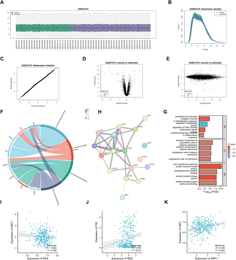
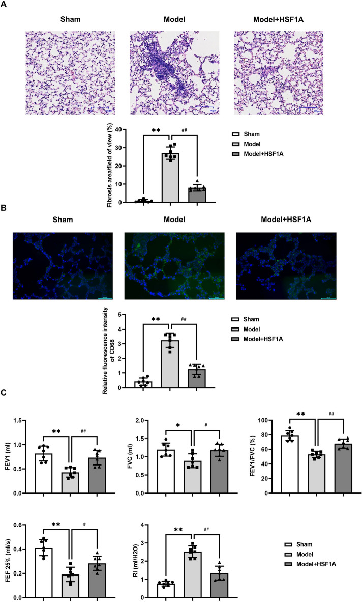
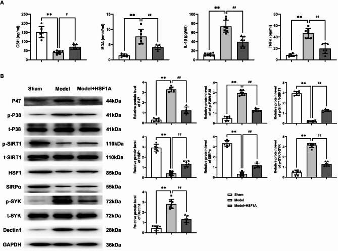
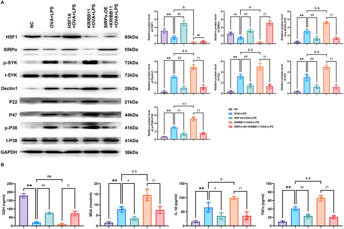
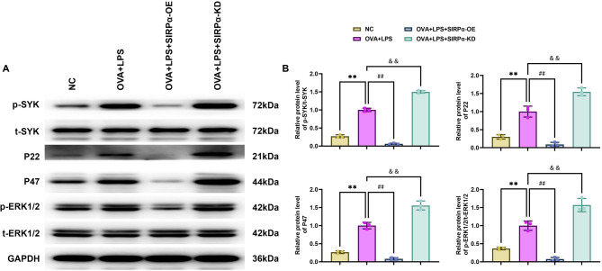
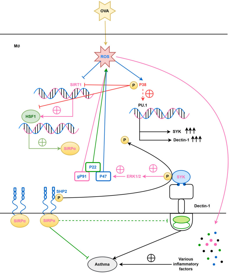
HSF1, SIRPα, and Dectin-1 play crucial roles in immune regulation and inflammatory responses, their rols in asthma remained unclear, thereby the study was carried on. Twenty-one SPF-grade C57BL/6 mice were randomly divided into three groups: sham group, Model group, and Model + HSF1A group, with seven mice in each group. Except for the sham group, the other two groups were induced with OVA to establish an asthma model. The Model + HSF1A group was additionally treated with HSF1A. General conditions of the mice were observed. Lung tissue damage was assessed with Masson staining. RAW264.7 cells were divided into NC group, OVA + LPS group, HSF1A + OVA + LPS group, KRIBB11 + OVA + LPS group, SIRPα-OE + KRIBB11 + OVA + LPS group, SIRPα-OE + OVA + LPS group and SIRPα-KD + KRIBB11 + OVA + LPS group. The levels of GSH, MDA, IL-1β and TNFα in serum and cell supernatants were determined by ELISA. Protein expression in lung tissue and RAW264.7 cells was detected by Western blotting. In in vivo experiments, OVA-induced asthmatic mice exhibited severe airway resistance, collagen deposition, and elevated ROS and pro-inflammatory cytokines.HSF1A treatment improved lung function, reduced fibrosis, and restored redox balance. In vitro, HSF1A enhanced SIRPα expression and inhibited SYK/Dectin-1 signaling in LPS/OVA-stimulated macrophages, whereas HSF1 knockdown exacerbated inflammation. overexpression of SIRPα reversed KRIBB11-induced SYK activation, confirming the regulatory role of HSF1. HSF1 in macrophages regulates ROS and inflammatory responses by modulating the SIRPα/Dectin-1/SYK balance, thereby inhibiting the progression of asthma.
Keywords: Asthma; HSF1; Inflammation; ROS; SIRPα/Dectin-1/SYK.
© 2025. The Author(s).
Conflict of interest statement
Competing interests: The authors declare no competing interests. Ethical approval: This study was supervised and approved by the Ethics Committee of the Second Hospital of Hebei Medical University. [Ethics Number: 2024-AE386]
Figures
Similar articles
-
IL-1β promotes IL-17A production of ILC3s to aggravate neutrophilic airway inflammation in mice.Immunology. 2025 Sep;176(1):16-32. doi: 10.1111/imm.13644. Epub 2023 Mar 29. Immunology. 2025. PMID: 36988516
-
miR-410 Regulates Helper T Cell Differentiation in Ovalbumin-Induced Asthma through the PI3K-AKT-VEGF Signaling Pathway.Int Arch Allergy Immunol. 2024;185(1):1-9. doi: 10.1159/000531493. Epub 2023 Sep 19. Int Arch Allergy Immunol. 2024. PMID: 37725935
-
Exosomes derived from baicalin-pretreated bone marrow mesenchymal stem cells inactivate the TLR4/MyD88/NF-kB pathway to improve asthma.Immunobiology. 2025 Jul;230(4):153103. doi: 10.1016/j.imbio.2025.153103. Epub 2025 Jul 16. Immunobiology. 2025. PMID: 40695068
-
Reactive Oxygen Species in Asthma: Regulators of Macrophage Polarization and Therapeutic Implications: A Narrative Review.J Asthma Allergy. 2025 Jul 25;18:1129-1146. doi: 10.2147/JAA.S529371. eCollection 2025. J Asthma Allergy. 2025. PMID: 40735422 Free PMC article. Review.
-
Anti-interleukin-13 and anti-interleukin-4 agents versus placebo, anti-interleukin-5 or anti-immunoglobulin-E agents, for people with asthma.Cochrane Database Syst Rev. 2021 Oct 19;10(10):CD012929. doi: 10.1002/14651858.CD012929.pub2. Cochrane Database Syst Rev. 2021. PMID: 34664263 Free PMC article.
References
LinkOut - more resources
Full Text Sources
Miscellaneous
