. 2025 Aug 13;16(1):7528.
doi: 10.1038/s41467-025-62741-7.

Multivalent interactions with CCR4-NOT and PABPC1 determine mRNA repression efficiency by tristetraprolin

Affiliations

Multivalent interactions with CCR4-NOT and PABPC1 determine mRNA repression efficiency by tristetraprolin

Filip Pekovic et al. Nat Commun. .

Abstract

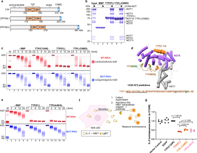
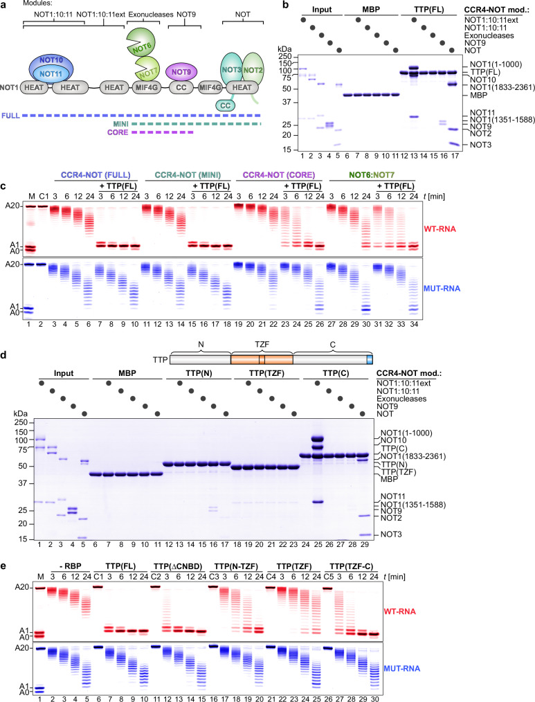
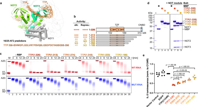
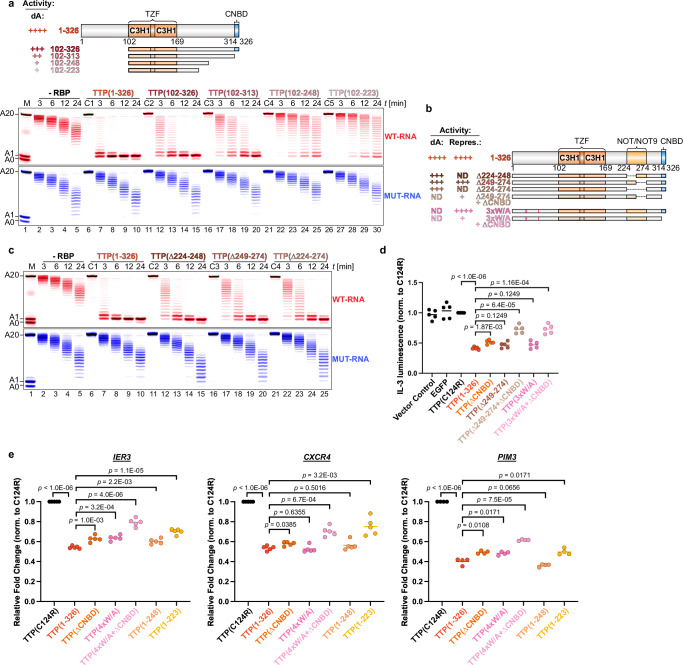
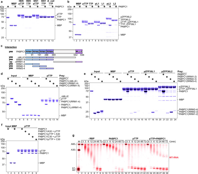
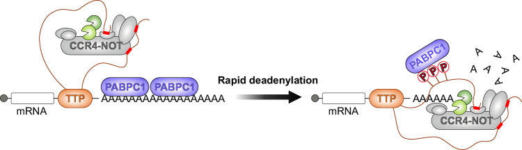
Tristetraprolin family of proteins regulate mRNA stability by binding to specific AU-rich elements in transcripts. This binding promotes the shortening of the mRNA poly(A) tail, or deadenylation, initiating mRNA degradation. The CCR4-NOT complex plays a central role in deadenylation, while the cytoplasmic poly(A)-binding protein PABPC1 typically protects mRNAs from decay. Here, we investigate how tristetraprolin interacts with CCR4-NOT and PABPC1 to control mRNA stability. Using purified proteins and in vitro assays, we find that tristetraprolin engages CCR4-NOT through multiple interaction sites and promotes its activity, emphasizing the importance of multivalent binding for efficient deadenylation. Phosphorylation of tristetraprolin does not affect its interaction with CCR4-NOT or its deadenylation activity, but is essential for tristetraprolin's binding to PABPC1. We propose that tristetraprolin promotes the processive deadenylation activity of CCR4-NOT on mRNAs containing AU-rich elements, with phosphorylation-dependent interactions with PABPC1 potentially enhancing deadenylation and promoting regulated mRNA decay.


Conflict of interest statement

Competing interests: The authors declare no competing interests.

Figures

Fig. 1
Fig. 1. Assessing the role of the C-terminal motif and tryptophan residues in TTP for CCR4–NOT recruitment and repression.
a Schematic representation of human tristetraprolin (TTP) and its human family members ZFP36L1 and ZFP36L2, highlighting the tandem zinc finger (TZF) domain necessary for RNA binding, containing two C3H1 zinc fingers, and the C-terminal NOT1-binding domain (CNBD). Also noted are the positions of the key tryptophan residues in TTP. b Pull-down assays comparing the interaction between CCR4–NOT (black circles) and either full-length TTP (FL) or TTP lacking the CNBD (ΔCNBD). Maltose-binding protein (MBP) was included as a negative control. Here and in all other figures, proteins were resolved by SDS-PAGE and visualized by Coomassie staining. c In vitro deadenylation assays demonstrating ARE-specific (WT-RNA, red, containing one TTP-binding motif; mutant RNA (MUT-RNA), blue) and TTP-dependent stimulation of deadenylation, compared with ΔCNBD and a non-RNA-binding mutant of TTP (C124R). Substrate RNAs (50 nM each) were incubated with 50 nM CCR4–NOT in the presence or absence (-RBP) of 100 nM TTP protein. Control reactions included RNA alone (C1) and RNA with the indicated TTP variants (C2–C4). Here and in all other figures, RNAs were resolved by urea-PAGE and visualized by fluorescence detection. The first lane here and in all following deadenylation assay gels indicates the size marker corresponding to the number of adenosines present. d AlphaFold2 predictions depicting fourteen out of twenty-five models of TTP interacting with the NOT9 module of CCR4–NOT. The first fourteen residues of TTP (orange) are predicted to bind the concave surface of NOT9 (purple), with tryptophan residues W32 (orange) and W38 (green) positioned near the tryptophan-binding pockets of NOT9. (e) Deadenylation assays as in (c), comparing full-length TTP (FL) to the TTP variant in which all tryptophan residues were mutated to alanine (4 × W/A). f Schematic illustration of the Nano-Glo HiBiT extracellular assay used to measure IL-3 protein production. Prepared in Affinity Designer 2. g Repression of IL-3 protein production by TTP variants. IL-3 luminescence was normalized to levels observed in cells expressing the non-RNA-binding mutant TTP(C124R). TTP(3×W/A) had tryptophan to alanine mutations at positions 32, 38, and 69. Statistical significance was determined using a two-tailed Student’s t-test followed by Holm-Šidák correction for multiple comparison and shown as exact p values. Each data point represents one biological replicate (n = 5).
Fig. 2
Fig. 2. TTP requires multiple CCR4–NOT interactions for efficient targeted deadenylation.
a Schematic representation of the human CCR4–NOT complex architecture, indicating the subcomplexes used: MINI (lacking the N-terminal portion of NOT1 and the NOT10/11 proteins) and CORE (lacking both the N- and C-terminal portions of NOT1 and the NOT2/3/10/11 proteins). Domains of NOT1 are denoted as follows: HEAT domain (α-helical HEAT-like repeats), MIF4G domain (‘middle of 4G’), and coiled-coil (CC) domain. Prepared in Affinity Designer 2. b Pull-down assays showing the interaction of full-length TTP (FL) with the indicated CCR4–NOT modules (black circles). MBP was included as a negative control. c Deadenylation assays comparing targeted deadenylation by full-length TTP (TTP-FL, 100 nM) in the presence of different CCR4–NOT subcomplexes. All CCR4–NOT subcomplexes were added at equimolar concentrations (50 nM) relative to substrate RNAs (50 nM), except for the exonuclease heterodimer, which was used at 250 nM. A control reaction containing only RNA and TTP (C1) was included. d Pull-down assays demonstrate that TTP’s N-terminal and C-terminal intrinsically disordered regions (IDRs) interact with multiple CCR4–NOT modules, as indicated (black circles). MBP was included as a negative control.The domain architecture of full-length TTP highlighting the N- and C-terminal IDRs surrounding the TZF domain is given for reference. The CNBD is included in the C-terminal IDR fragment. e Deadenylation assays comparing the stimulation of deadenylation by TTP (FL) with ∆CNDB and TTP variants in which either IDR was fused to the TZF RNA-binding domain. All TTP variants were added at 100 nM, in twofold excess over CCR4–NOT (50 nM) and substrate RNAs (50 nM). Controls included reactions containing only RNA and the indicated TTP variant (C1-C5).
Fig. 3
Fig. 3. The C-terminal unstructured region of TTP is essential for interaction with the NOT module and efficient deadenylation.
a Alignment of sixteen out of twenty-five AlphaFold2 predictions reveals a potential interaction surface between TTP (orange) and the NOT module (NOT1 in grey, NOT2 in light green, NOT3 in dark green). A short region within the C-terminal IDR of TTP converges around NOT3. b Domain organization and summary of activity for the C-terminal truncations of TTP used in deadenylation assays (labeled ‘dA’; panel c) and protein production assays (labeled ‘Repres.’; panel e). c Deadenylation assays assessing the effect of gradual C-terminal truncations of TTP on targeted deadenylation. All TTP variants were added at 100 nM, in twofold excess over CCR4–NOT (50 nM) and substrate RNAs (50 nM). Control reactions included reactions with RNA and the indicated TTP variant (C1-C6) alone. d Pull-down assays with the NOT module and the indicated TTP fragments (colored circles). MBP (black circle) was included as a negative control. e Effect of C-terminal IDR truncations of TTP on the repression of IL-3 protein production in co-transfection assays, measured by relative luminescence. Statistical significance was determined using a two-tailed Student’s t-test followed by Holm-Šidák correction for multiple comparison and shown as exact p values. Each data point represents one biological replicate (n = 5).
Fig. 4
Fig. 4. Multivalent interactions between TTP and CCR4–NOT are crucial for rapid deadenylation and repression.
a Deadenylation assays assessing the ability of TTP fragments lacking both the N-terminal IDR and the indicated regions within the C-terminal IDR to stimulate targeted deadenylation. Domain organization schematic and associated deadenylation (‘dA’) activity summary is presented. All TTP proteins were added at 100 nM, in twofold excess over CCR4–NOT (50 nM) and substrate RNA (50 nM). Controls included reactions with RNA and the indicated TTP variant (C1–C5) alone. b Domain organization and summary of activity for TTP deletion and mutant variants used in deadenylation assays (c) and in co-transfection protein production assays (panel d). Mutated tryptophan residues (W32A/W38A/W69A) are indicated with pink bars. c Deadenylation assays testing deletion variants of the region in TTP that interacts with the NOT9 and NOT modules, assessing their ability to promote targeted deadenylation. The assay was performed as in (a). d The effect of deleting two critical CCR4–NOT-interacting regions in TTP on the repression of IL-3 protein production was measured via secreted IL-3 luminescence. Either both the CNBD and part of the C-terminal repressor region (∆249–274) were deleted (light brown) or the CNBD was deleted in combination with point mutations of the three tryptophan residues (W32A/W38A/W69A) in TTP’s N-terminal IDR (light pink). Each data point represents one biological replicate (n = 5). e RT-qPCR of endogenous IER3, CXCR4, and PIM3 in HEK-293T cells transduced with CMV-driven vectors expressing various TTP variants: wild-type (1–326), RNA-binding mutant (C124R), ∆CNBD, all tryptophan residues to alanine mutant (4xW/A), a combination of ∆CNBD and 4xW/A, and C-terminal truncations (1–248 and 1–223). Each data point represents one biological replicate (n = 5 for IER3 and CXCR4, n = 4 for PIM3). Statistical significance in (d, e) was determined using a two-tailed Student’s t-test followed by Holm-Šidák correction for multiple comparison and shown as exact p values.
Fig. 5
Fig. 5. Phosphorylation of TTP and its family members does not directly affect CCR4–NOT interaction or deadenylation activity.
a Phosphorylated TTP (pTTP) and its human family members (pZFP36L1, pZFP36L2) were expressed and isolated from eukaryotic systems (human HEK-293 and insect Sf21 cells). Phosphorylation status was confirmed by treating proteins with protein phosphatase (black circles) and comparing migration patterns to untreated samples via SDS-PAGE. TTP expressed in E. coli served as a non-phosphorylated control. b Diagram comparing previously reported phosphorylation sites in TTP based on the PhosphoSitePlus database to sites identified by mass spectrometry in TTP isolated from HEK-293 or Sf21 cells in this study. Overlapping sites (orange triangle), non-overlapping sites (red circles) and new sites (green rectangles) are indicated. c Deadenylation assays testing the activity of pTTP (expressed in HEK-293 or Sf21 cells) and phosphorylated pZFP36L1 and pZFP36L2 in targeted deadenylation. The activity of pTTP was also compared to non-phosphorylated TTP. All TTP proteins were used at 100 nM, in twofold excess over CCR4–NOT (50 nM) and substrate RNA (50 nM). Controls included reactions with TTP and RNA alone (C1–C5). d Pull-down assays demonstrating the interactions between phosphorylated TTP, pZFP36L1, and pZFP36L2, with CCR4–NOT modules (black circles). MBP was included as a negative control.
Fig. 6
Fig. 6. Phosphorylation-dependent interaction between TTP and PABPC1 promotes CCR4–NOT-mediated shortening of PABPC1-coated poly(A) tails.
a, b Pull-down assays testing the interaction between the cytoplasmic poly(A)-binding protein (PABPC1) and either phosphorylated TTP (pTTP) expressed in HEK-293, Sf21, or E. coli cells, or phosphorylated human family members. Proteins were incubated with or without protein phosphatase (black circles) to assess the effect of dephosphorylation on the interaction. MBP was included as a negative control. c Domain architecture of PABPC1 and a summary of observed interactions between PABPC1 fragments and pTTP (d, e). RRM: RNA recognition motif; MLLE: mademoiselle protein–protein interaction domain. d Pull-down assays showing the interaction between pTTP and various fragments of PABPC1, as indicated (black circles). MBP was included as a negative control. e Characterization of the interaction between the RRMs of PABPC1 and pTTP, pZFP36L1, and pZFP36L2, via pull-down assays. f Assessment of PABPC’s ability to interact simultaneously with a poly(A) tail and pTTP. Complexes of PABPC1 with a poly(A) tail of 30 adenosine residues (PABPC1/A30) were preformed before pTTP addition, or complexes of PABPC1 with pTTP (PABPC1/pTTP) were formed before adding A30 oligo. To confirm the poly(A) specificity, incubations were carried out with a C30 oligo. MBP was included as a negative control. g Deadenylation assay testing the effect of PABPC1 on targeted deadenylation by pTTP. A PABPC1:RNA complex was preformed first by addition of PABPC (150 nM) in threefold excess over substrate RNA (50 nM) to saturate the poly(A) tail. pTTP protein (100 nM) was added in twofold excess over CCR4–NOT (50 nM) and substrate RNA (50 nM). Controls included reactions lacking pTTP or PABPC1(-RBP), and reactions with only RNA and PABPC1 (C2) or pTTP (C3) or PABPC1 and pTTP (C4).
Fig. 7
Fig. 7. A proposed model of TTP-mediated rapid deadenylation via multivalent interactions with CCR4–NOT and PABPC1.
Schematic model illustrating how TTP exploits multivalent interactions with two key regulatory effectors of mRNA fate — CCR4–NOT and PABPC1. Short segments within TTP’s intrinsically disordered C-terminal region (red rectangles) serve as docking sites that scaffold these effectors and direct rapid, transcript-specific deadenylation. Phosphorylation of TTP (red “lollipop” symbols) promotes high-affinity binding to PABPC1, displacing PABPC1 from the poly(A) tail and thereby clearing access for CCR4–NOT to engage the tail and catalyze efficient deadenylation. Prepared in Affinity Designer 2.

Similar articles

References

    1. Sanduja, S., Blanco, F. F. & Dixon, D. A. The roles of TTP and BRF proteins in regulated mRNA decay: TTP and BRF proteins in regulated mRNA decay. Wiley Interdiscip. Rev. RNA2, 42–57 (2011). - PMC - PubMed
    1. Blackshear, P. J. & Perera, L. Phylogenetic distribution and evolution of the linked RNA-binding and NOT1-binding domains in the tristetraprolin family of tandem CCCH zinc finger proteins. J. Interferon Cytokine Res.34, 297–306 (2014). - PMC - PubMed
    1. Wells, M. L., Perera, L. & Blackshear, P. J. An ancient family of RNA-binding proteins: Still important!. Trends Biochem. Sci.42, 285–296 (2017). - PMC - PubMed
    1. Lai, W. S., Wells, M. L., Perera, L. & Blackshear, P. J. The tandem zinc finger RNA binding domain of members of the tristetraprolin protein family. Wiley Interdiscip. Rev. RNA10, e1531 (2019). - PMC - PubMed
    1. Khabar, K. S. A. Post-transcriptional control during chronic inflammation and cancer: A focus on AU-rich elements. Cell. Mol. Life Sci.67, 2937–2955 (2010). - PMC - PubMed

LinkOut - more resources