. 2025 Aug 13;8(1):1209.
doi: 10.1038/s42003-025-08613-8.

rTMS ameliorates cerebral ischemia-reperfusion injury by inhibiting Golgi apparatus stress through epigenetic modulation of Gli2

Affiliations

rTMS ameliorates cerebral ischemia-reperfusion injury by inhibiting Golgi apparatus stress through epigenetic modulation of Gli2

Chunjiao Zhu et al. Commun Biol. .

Abstract

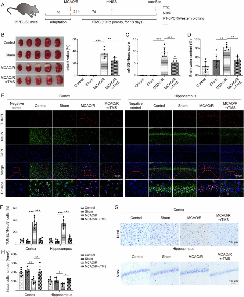
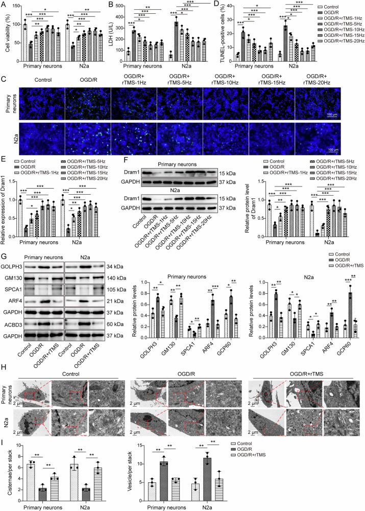
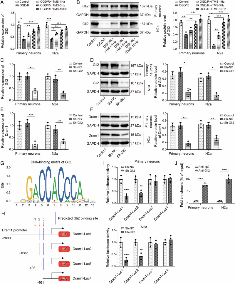
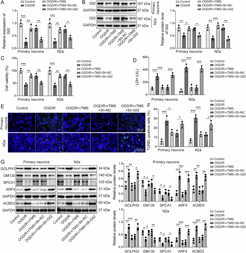
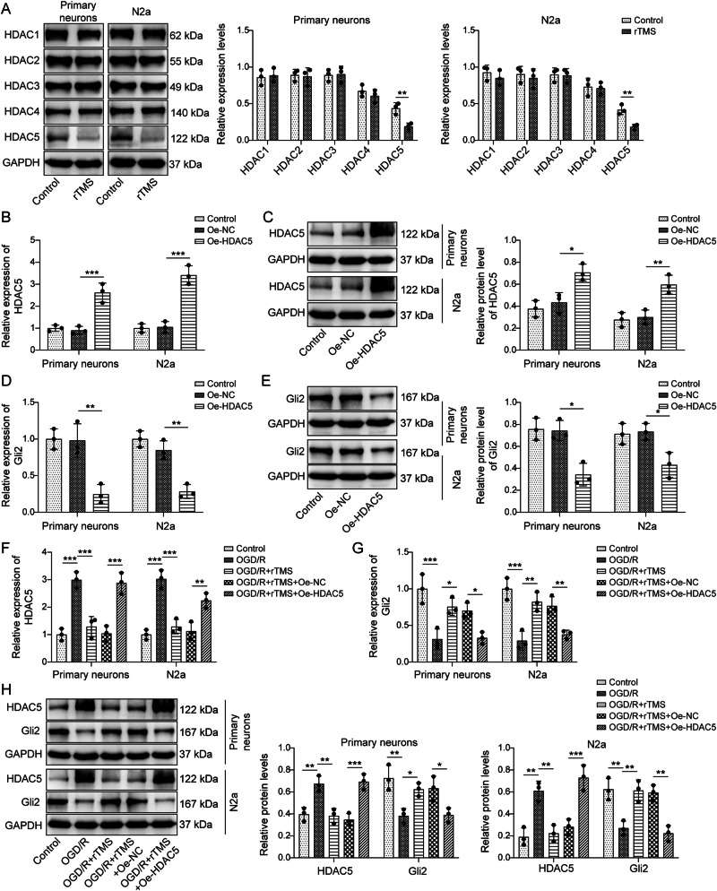
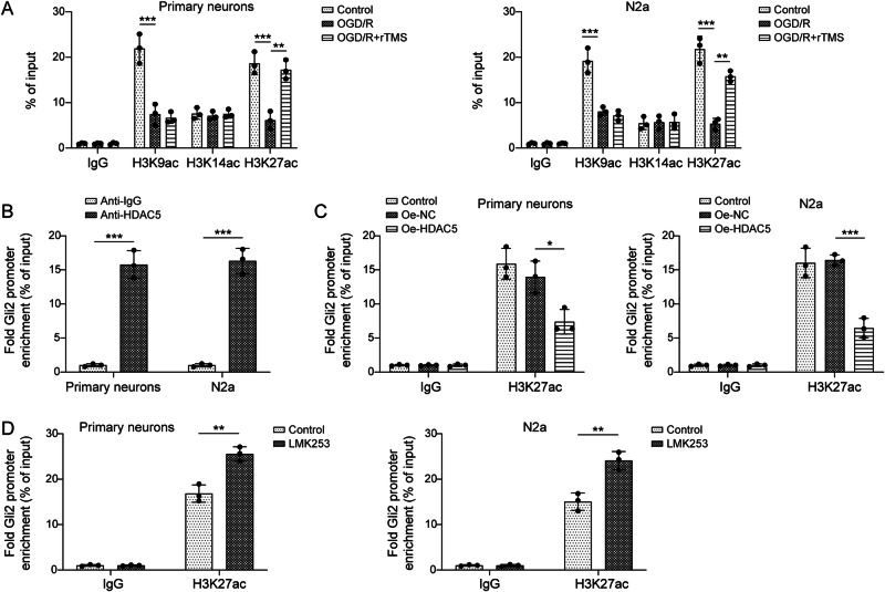
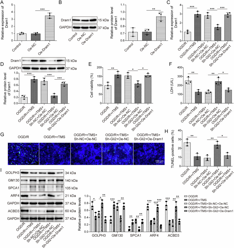
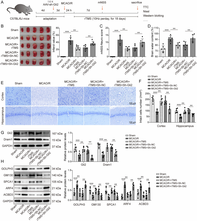
Cerebral ischemic stroke represents a primary cause of permanent disability and mortality globally. Repetitive transcranial magnetic stimulation (rTMS) has emerged as a prominent focus in treating a wide range of neurological disorders. In this study, we explore the role of rTMS in alleviating cerebral ischemia-reperfusion (I/R) injury by mediating Golgi apparatus (GA) stress. Here, we find that rTMS upregulates Dram1 expression and ameliorates GA stress in cerebral I/R injury in vivo and in vitro. Gli2 transcriptionally activates Dram1. HDAC5 inhibits H3K27ac modification of Gli2 promoter. rTMS promotes Gli2 expression by inhibiting HDAC5. Gli2 knockdown reverses the inhibitory effect of rTMS on OGD/R-induced neuronal GA stress. In conclusion, rTMS inhibits HDAC5-mediated deacetylation of Gli2 promoter to promote the transcriptional activation of Dram1, thereby suppressing cerebral I/R-induced GA stress. Targeting Gli2/ Dram1 axis may be an effective way to enhance the anti-ischemic stroke effect of rTMS.


Conflict of interest statement

Competing interests: The authors declare no competing interests.

Figures

Fig. 1
Fig. 1. rTMS ameliorated brain injury in MCAO/R mice.
C57BL/6 J mice were treated with MCAO/R surgery to induce cerebral I/R injury, followed by 10 Hz rTMS stimulation. A The flow chart of experiments with MCAO/R treatment and rTMS stimulation in mice. B TTC staining was performed to test the infarct area in mouse brain tissues at 18 days after rTMS treatment (n = 6). C Neurological function in mice at 7 days after rTMS treatment was assessed using mNSS (n = 6). D The water content of the mouse brain was determined using the wet/dry weight ratio method (n = 6). E, F Colocalization of NeuN and TUNEL in cortical and hippocampal regions of mouse brain tissues (n = 6). G, H Neurological damage in cortex and hippocampus was examined by Nissl’s staining (n = 6). For (BH), one-way ANOVA with Tukey’s post hoc test was performed to analyze data. *P < 0.05, **P < 0.01 and ***P < 0.001.
Fig. 2
Fig. 2. rTMS suppressed OGD/R-induced GA stress and neuronal damage by upregulating Dram1.
Primary neurons and N2a cells were subjected to OGD/R treatment and treated with 1, 5, 10, 15 and 20 Hz rTMS, respectively. A CCK-8 assay was employed to measure cell viability. B LDH levels were tested in primary neurons and N2a cells by ELISA kit. C, D Apoptosis was evaluated by TUNEL staining. E, F Dram1 mRNA and protein levels were detected using RT-qPCR and western blotting, respectively. Based on the experimental results, the optimal rTMS frequency (10 Hz) was selected for subsequent experiments. G Levels of GA functional and structural proteins (GOLPH3, GM130, SPCA1, ARF4, and ACBD3) were measured by western blotting. H, I Morphology and ultrastructure of GA in primary neurons and N2a cells were detected using transmission electron microscopy and the numbers of cisternae and vesicle surrounding each stack were analyzed. n = 3. For AI, one-way ANOVA with Tukey’s post hoc test was performed to analyze data. *P < 0.05, **P < 0.01 and ***P < 0.001.
Fig. 3
Fig. 3. Dram1 was transcriptionally activated by Gli2 in neurons.
A, B Primary neurons and N2a cells were subjected to OGD/R treatment for 3 h, and then cells were stimulated using 1, 5, 10 Hz rTMS. Gli2 mRNA and protein levels were examined by RT-qPCR and western blotting. CF sh-NC or sh-Gli2 was transfected into primary neurons and N2a cells, respectively, and the levels of Gli2 (C, D) and Dram1 (E, F) were examined by RT-qPCR and western blotting, respectively. G The conserved DNA-binding motifs of Gli2 were predicted by JASPAR database (https://jaspar.elixir.no/). H The truncated image of the Dram1 promoter region. I The relative luciferase activities of Dram1-Luc-1, Dram1-Luc-2, Dram1 Luc-3, and Dram1-Luc4 was tested by dual-luciferase reporter gene experiments in primary neurons and N2a cells after knockdown of Gli2. J The binding relationship of Gli2 to the Dram1 promoter was verified by ChIP. n = 3. One-way ANOVA with Tukey’s post hoc test (for AF) and Student’s t test (for I, J) were performed to analyze data. *P < 0.05, **P < 0.01 and ***P < 0.001.
Fig. 4
Fig. 4. Knockdown of Gli2 reversed the effects of rTMS on OGD/R-induced GA stress and neuronal damage.
The sh-NC or sh-Gli2 was transfected into primary neurons and N2a cells treated with OGD/R and rTMS, respectively. A, B RT-qPCR and western blotting assays were performed to determine the Gli2 levels in primary neurons and N2a cells. C Cell viability was assayed using a CCK-8 kit. D LDH levels in primary neurons and N2a cells were measured using ELISA kit. E, F Apoptosis was examined using the TUNEL assay. G The expression levels of GA functional and structural proteins (GOLPH3, GM130, SPCA1, ARF4, and ACBD3) were determined by western blotting. n = 3. One-way ANOVA with Tukey’s post hoc test was performed to analyze data. *P < 0.05, **P < 0.01 and ***P < 0.001.
Fig. 5
Fig. 5. rTMS promoted Gli2 expression by inhibiting HDAC5.
A HDAC1-5 expression levels in rTMS-treated primary neurons and N2a cells were checked by western blotting. B, C The effect of HDAC5 overexpression was detected by RT-qPCR and western blotting in primary neurons and N2a cells. D, E RT-qPCR and western blotting were applied to check the influence of HDAC5 overexpression on Gli2 mRNA and protein levels. FH HDAC5 overexpression vector or its negative control oe-NC was transfected into primary neurons and N2a cells with OGD/R and rTMS treatments, and mRNA (F, G) and protein (H) levels of HDAC5 and Gli2 were detected using RT-qPCR and western blotting. n = 3. Student’s t test (for A) and one-way ANOVA with Tukey’s post hoc test (for BH) were performed to analyze data. *P < 0.05, **P < 0.01 and ***P < 0.001.
Fig. 6
Fig. 6. HDAC5 inhibited H3K27ac modification of the Gli2 promoter region in neurons.
A Primary neurons and N2a cells were treated with OGD/R and 10 Hz rTMS. H3K9ac, H3K14ac, and H3K27ac enrichment in Gli2 promoter region was detected by ChIP assay. B ChIP was performed to assay the binding relationship between HDAC5 and Gli2 promoter in primary neurons and N2a cells. C The effect of overexpression of HDAC5 on H3K27ac enrichment in Gli2 promoter region was tested by ChIP assay. D H3K27ac enrichment in Gli2 promoter region was measured by ChIP after treatment of primary neurons and N2a cells with an inhibitor of HDAC5 (LMK253). n = 3. One-way ANOVA with Tukey’s post hoc test (for A and C) and student’s t test (for B, D) were performed to analyze data. *P < 0.05, **P < 0.01 and ***P < 0.001.
Fig. 7
Fig. 7. Overexpression of Dram1 reversed the effects of Gli2 knockdown on OGD/R-induced neuronal GA stress and damage under rTMS treatment.
A, B After Dram1 overexpression vector or its negative control oe-NC was transfected into N2a cells, Dram1 levels were tested by RT-qPCR and western blotting. sh-Gli2, Dram1 overexpression vector, and their negative controls were transfected into N2a cells, which were then subjected to OGD/R and 10 Hz rTMS treatment. C, D RT-qPCR and western blotting were applied to analyze the expression levels of Dram1. E Cell viability was determined by the CCK-8 assay. F LDH levels were tested using ELISA kit. G, H Apoptosis was assessed by TUNEL staining. I Levels of GOLPH3, GM130, SPCA1, ARF4, and ACBD3 were assayed using western blotting. n = 3. One-way ANOVA with Tukey’s post hoc test was performed to analyze data. *P < 0.05, **P < 0.01 and ***P < 0.001.
Fig. 8
Fig. 8. Knockdown of Gli2 reversed the effects of rTMS in ameliorating GA stress and brain injury in MCAO/R mice.
The sh-Gli2 or sh-NC adenovirus was injected into the lateral ventricle of MCAO/R mice, which were then treated with 10 Hz rTMS. A The flow chart of the rTMS and adenovirus treatment to C57BL/6 J mice. B TTC staining was performed to test the infarct area in the mouse brain tissues at 18 days after rTMS treatment (n = 6). C Neurological function in mice was assessed using mNSS at 7 days after rTMS treatment (n = 6). D The water content of the mouse brain was determined using the wet/dry weight ratio method (n = 6). E, F Nissl staining was used to measure nerve damage in mice (n = 6). G The protein levels of Gli2 and Dram1 in brain tissues were determined by western blotting assays (n = 6). H The protein levels of GA functional and structural proteins (GOLPH3, GM130, SPCA1, ARF4, and ACBD3) in brain tissues were measured using western blotting (n = 6). One-way ANOVA with Tukey’s post hoc test was performed to analyze data. *P < 0.05, **P < 0.01 and ***P < 0.001.

Similar articles

References

    1. Mao, Z. et al. Ligustilide ameliorates hippocampal neuronal injury after cerebral ischemia reperfusion through activating PINK1/Parkin-dependent mitophagy. Phytomedicine101, 154111 (2022). - PubMed
    1. Chen, Y. et al. Srs11-92, a ferrostatin-1 analog, improves oxidative stress and neuroinflammation via Nrf2 signal following cerebral ischemia/reperfusion injury. CNS Neurosci. Ther.29, 1667–1677 (2023). - PMC - PubMed
    1. Lan, Z. et al. Curcumin-primed olfactory mucosa-derived mesenchymal stem cells mitigate cerebral ischemia/reperfusion injury-induced neuronal PANoptosis by modulating microglial polarization. Phytomedicine129, 155635 (2024). - PubMed
    1. Xu, D. et al. Orexin-A alleviates astrocytic apoptosis and inflammation via inhibiting OX1R-mediated NF-κB and MAPK signaling pathways in cerebral ischemia/reperfusion injury. Biochim Biophys. Acta Mol. Basis Dis.1867, 166230 (2021). - PubMed
    1. Huang, Y. et al. UBIAD1 alleviates ferroptotic neuronal death by enhancing antioxidative capacity by cooperatively restoring impaired mitochondria and Golgi apparatus upon cerebral ischemic/reperfusion insult. Cell Biosci.12, 42 (2022). - PMC - PubMed

LinkOut - more resources