. 2025 Aug 13;25(1):304.
doi: 10.1186/s12906-025-05053-z.

Urtica dioica leaf infusion enhances cisplatin-induced apoptosis in ovarian cancer cells in vitro

Affiliations

Urtica dioica leaf infusion enhances cisplatin-induced apoptosis in ovarian cancer cells in vitro

Perla Nafeh et al. BMC Complement Med Ther. .

Abstract

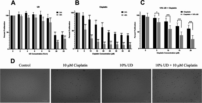
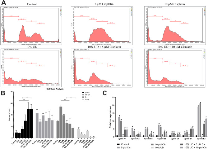
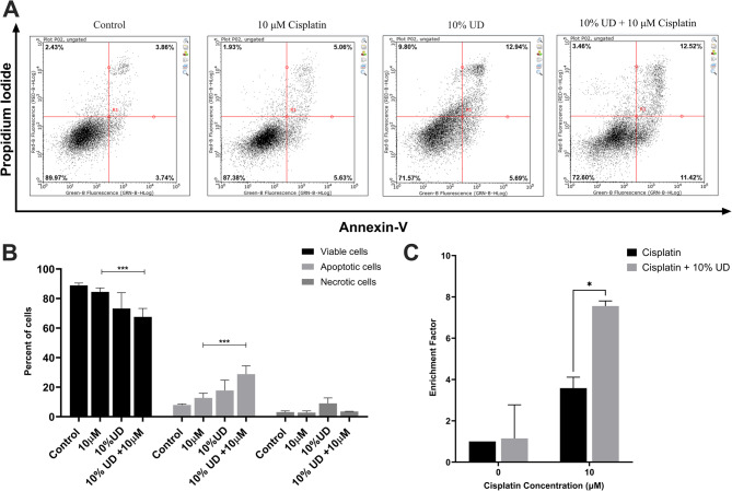
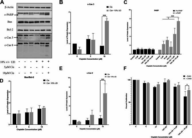
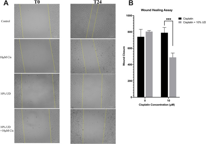
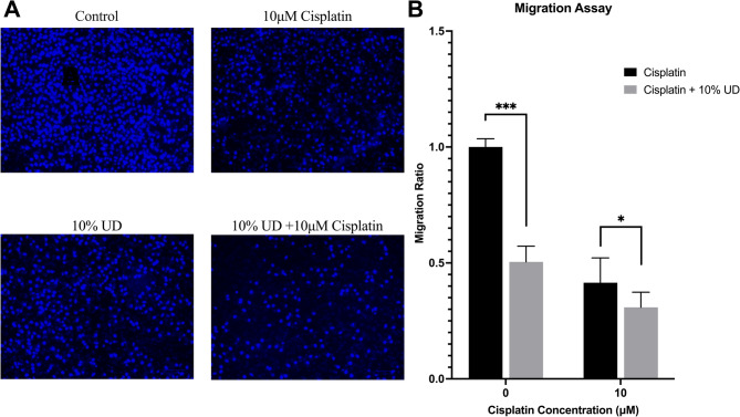
Urtica Dioica (UD) or stinging nettle has been widely used for its therapeutic benefits and biological activities. Recent studies have reported its beneficial effect in treating cancer, most importantly when combined with chemotherapeutic drugs. To our knowledge, no studies investigated the potential effect of UD to enhance the sensitivity of ovarian cancer cells to cisplatin. In this study we aim to investigate whether this combination might possess anti-proliferative, pro-apoptotic, and anti-metastatic properties on one of the most aggressive ovarian cancer cell lines, SKOV-3 cells, in vitro. To elucidate its therapeutic values, cytotoxicity and DNA fragmentation assays were performed along with cell cycle and apoptosis assays using flow cytometry, RT-qPCR, and western blot analysis. Additionally, wound healing and trans-well migration assays were used to study the effect of this combination on the motility of ovarian cancer cells. Results showed that the combination of nettle extract and cisplatin significantly decreased the proliferation of SKOV-3 cells in a dose and time-dependent manner compared to each treatment alone by inducing cellular death as revealed by major apoptotic markers including phosphatidylserine translocation to the outer membrane leaflet, DNA fragmentation, and the upregulation of cleaved PARP protein. Further evaluation verified the activation of extrinsic apoptosis via the caspase-dependent pathway as demonstrated by the upregulated expression levels of caspases 3 and 8. Finally, the combination of nettle tea and cisplatin showed an inhibitory effect on the motility and migratory capacities of SKOV-3 cells. As a result, Urtica Dioica leaf infusion was found effective in enhancing cisplatin-induced apoptosis while inhibiting the tumor progression of one of the most aggressive ovarian cancer cells in vitro.

Keywords: Urtica dioica; Apoptosis; Cisplatin; Combination therapy; Motility; Ovarian cancer.


Conflict of interest statement

Declarations. Ethics approval and consent to participate: No human subjects were used in this study. Consent for publication: Plant sampling was collected on a public land in Bawarji, Lebanon, which does not require Lebanese government permission. Competing interests: The authors declare no competing interests.

Figures

Fig. 1
Fig. 1
MTS Cytotoxicity Assay. A Percent proliferation of the SKOV-3 cells after 24 h and 48 h of treatment with different concentrations of UD (0–25% v/v). Results are compared to untreated negative control cells. Significant growth inhibition was observed upon UD treatment (p < 0.001).(B Percent proliferation of the SKOV-3 cells after 24 h and 48 h of treatment with different concentrations of cisplatin (0-40µM). Results are compared to untreated negative control cells and a significant decrease in proliferation was observed upon increasing the cisplatin concentration (p < 0.001). C Percent proliferation of the SKOV-3 cells after 24 h upon co-treatment of increasing concentrations of cisplatin (0-20µM) with 10% v/v UD. Results are compared to those treated with cisplatin alone. Each value represents the mean ± SD from three separate experiments; statistical analysis was determined by using Student’s t-test with * p < 0.05, ** p < 0.01, and *** p < 0.001. D Brightfield images using the ZOE Fluorescent Microscope showing the shape and physical state of the cells 24 h after their respective treatment, where dead cells are highlighted with arrows
Fig. 2
Fig. 2
A Cell cycle analysis of SKOV-3 cells treated with cisplatin (5µM, 10µM) and UD (10% v/v), individually or in combination for 24 h. B A significant dose dependent increase in the pre-G phase and a significant decrease in the S phase, reported after 24 h (p < 0.001). C Relative expression of cyclins A1, A2, B1, B2, D1, E1 and E2 by RT-qPCR in SKOV-3 cells treated with cisplatin (5µM, 10µM), 10% v/v UD, and their combinations. GAPDH was used as a reference gene. Results are compared to cells treated with cisplatin alone or to the untreated control cells. The data is represented as mean ± SD from three separate experiments. Statistical significance is reported as *** corresponding p-value < 0.001, “a” corresponding to the significance relative to the control, and “b” the significance relative to 5µM and 10µM cisplatin
Fig. 3
Fig. 3
Representative fluorescent microscopy images showing Annexin V staining of SKOV-3 cells treated for 24 h with either 10µM cisplatin, 10% v/v UD, or their combination. A gradual increase in the number of Annexin-positive cells can be observed after 24 h of treatment, whereby most cells become positively stained at 10µM cisplatin combined with 10% v/v UD
Fig. 4
Fig. 4
Annexin V/PI Dual Staining of SKOV-3 cells. A Dot plot analysis using flow cytometry of SKOV-3 cells treated with 10µM cisplatin or 10% v/v UD or co-treatment of 10% v/v UD with 10µM cisplatin for 24 h.B Histogram representing the percentage of viable and apoptotic cells showing a significant increase in apoptotic cells coupled with a significant decrease in viable cells (p < 0.001). Results are compared to cells treated with cisplatin alone or to the untreated control cells. The data is represented as mean ± SD from three separate experiments. Statistical significance is reported as *** corresponding p-value < 0.001. C Cell Death detection ELISA. Quantification of DNA fragmentation in SKOV-3 cells upon treatment with 10µM cisplatin, 10% v/v UD alone, and their combination for 24 h. A significant increase in DNA fragmentation was observed in SKOV-3 cells after 24 h of treatment (p < 0.05). Results are compared to cells treated with cisplatin alone or to the untreated control cells. The data is represented as mean ± SD from three separate experiments. Statistical significance is reported as * corresponding p-value < 0.05
Fig. 5
Fig. 5
Western Blot analysis showing the combination of cisplatin (5 and 10µM) and UD (10%v/v) on SKOV-3 cells after 24 h of treatment. A Western blots of apoptotic proteins. The protein expression level of B cleaved-caspase 3 (c-cas3), C PARP, D Bax and Bcl2, and E cleaved-caspase 8 (c-cas8) was quantified using ImageJ software. A significant increase in c-PARP, c-cas3 and c-cas8 (p < 0.001) was observed after 24 h of treatment. F MTS cell viability assay was performed following treatment for 24 h with cisplatin (5µM, 10µM), UD (10% v/v), and the combination of cisplatin and UD with or without the caspase inhibitor ZVAD. Reversal of cell death was observed upon the addition of the caspase inhibitor to the cells (p < 0.05). The data are represented as mean ± SD from three separate experiments. Statistical significance is reported as * corresponding to p-value < 0.05 and *** to p-value < 0.001
Fig. 6
Fig. 6
Wound healing assay of SKOV-3 cells treated with either 10µM cisplatin, 10% v/v UD, or their combination. A Brightfield microscopic images captured at T = 0 and T = 24 h. B Quantification of the wound closure using ImageJ software. A significant decrease in wound closure was observed in cells treated with UD and cisplatin as compared to the cisplatin treatment alone (p < 0.001). The data are represented as mean ± SD from three separate experiments. Statistical significance is reported as * corresponding to p-value < 0.05 and *** to p-value < 0.001
Fig. 7
Fig. 7
Trans well migration assay on SKOV-3 cells treated with cisplatin (10µM), UD (10%V/V) and their combination for 24 h. A Fluorescent microscopic images of migratory SKOV-3 cells after 24 h of treatment. B Quantitative assessment of migratory cells using ImageJ software. A significant decrease in the number and percentage of migrated SKOV-3 cells through the porous membrane was observed upon exposure to UD and cisplatin (p < 0.001). The data are represented as mean ± SD from three separate experiments. Statistical significance is reported as * corresponding to p-value < 0.05 and *** to p-value < 0.001

Similar articles

References

    1. Yousefi M, Dehghani S, Nosrati R, Ghanei M, Salmaninejad A, Rajaie S, et al. Current insights into the metastasis of epithelial ovarian cancer - hopes and hurdles. Cell Oncol. 2020;43(4):515–38. - PubMed
    1. Ovarian Cancer Research Alliance. Ovarian Cancer Statistics. https://ocrahope.org/for-patients/gynecologic-cancers/ovarian-cancer/ova.... Accessed 20 Nov 2024.
    1. Bhatt A, Glehen O. The role of cytoreductive surgery and hyperthermic intraperitoneal chemotherapy (HIPEC) in ovarian cancer: A review. Indian J Surg Oncol. 2016;7(2):188–97. - PMC - PubMed
    1. Leong E, Ong SK, Jali F, Naing L. Incidence, mortality and survival analysis of epithelial ovarian cancer in Brunei Darussalam. 2022. https://www.researchsquare.com/article/rs-954088/v2. Accessed 25 Oct 2024. - PMC - PubMed
    1. Zhang M, Yin R, Li K. Advances and challenges in the origin and evolution of ovarian cancer organoids. Front Oncol. 2024;14:1429141. - PMC - PubMed