Review
. 2025 Aug 13;23(1):906.
doi: 10.1186/s12967-025-06919-z.

From network biology to immunity: potential longitudinal biomarkers for targeting the network topology of the HIV reservoir

Affiliations
Review

From network biology to immunity: potential longitudinal biomarkers for targeting the network topology of the HIV reservoir

Heng-Chang Chen. J Transl Med. .

Abstract

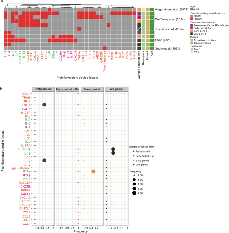
In the "omics" era, studies often utilize large-scale datasets, eliciting the overall functional machinery of a network's organization. In this context, determining how to read the enormous number of interactions in a network is imperative to comprehend its functional organization. Topology is the principal attribute of any network; as such, topological properties help to elucidate the roles of entities and represent a network's behavior. In this review, I showcase the foundational concepts involved in graph theory, which form the basis of network biology, and exemplify the application of this conceptual framework to bridge the connection between the task-evoked functional genome network of the HIV reservoir. Furthermore, I point out potential longitudinal biomarkers identified using network-based analysis and systematically compare them with other potential biomarkers identified based on experimental research with longitudinal clinical samples.

Keywords: Biological networks; Deep learning; Elite controllers; Graph theory; HIV reservoir; Immunologic signatures; Longitudinal biomarkers; Network analysis; Network topology.


Conflict of interest statement

Declarations. Ethics approval and consent to participate: Not applicable. Consent for publication: Not applicable. Competing interests: The author declares that they have no competing interests.

Figures

Fig. 1
Fig. 1
Schematic illustration of different forms of a graph. a A simple graph. It is composed of vertices (V) and edges (E). A V set includes the vertices 1, 2, 3, and 4; an E set includes the edges 12, 13, 14, 23, 24, and 34. b A directed graph. Directions, represented by arrowheads, were assigned to the edges. c A disconnected graph. No path is connected between a V set, 1, 2, 3, and 4, and another set, 5, 6, and 7. d A planar graph. e A forest, f A tree. g A simple graph, illustrating a walk, a path, and a cycle. h A bipartite graph that consists of two disjoint V sets, X labeled in blue and Y labeled in magenta. Of note, definitions, and descriptions corresponding to each form of the graph were detailed in the main text and the Glossary. i Schematic illustration of the hypothesis of the topological network representing HIV reservoirs. In our studies, we hypothesize that HIV-targeted genes (represented by red triangles shown in the cartoon) harboring similar biological functions related to immunity can form different gene sets, namely “immunologic signatures”, to satisfy the need for immunity at different stages of HIV infections. Such signatures can form a task-evoked network representing HIV reservoirs at a global level of network organization. Circles in blue and magenta represent different immunologic signatures (serve as vertices) assigned to various tasks, thus clustering into disproportionate blocks of modules. Edges were calculated based on correlation coefficients between two adjacent vertices
Fig. 2
Fig. 2
Schematic representation of the evolution of the graph network, representing the configuration of HIV reservoirs alongside HIV infections associated with ART and that structured in elite controllers. As computing the Pearson distance between the graph networks from reservoirs in pretreatment HIV-infected individuals, patients subjected to short and long periods of ART (graph networks highlighted in green circles), and elite controllers (the graph network highlighted in a pink circle), it is observed that the graph network between patients subjected to a long period of ART and elite controllers demonstrated the farthest graph distance, suggesting that a lack in the graph isomorphism of the networks between these two types of HIV-infected individuals. More details can be referred to as Wiśniewski et al. [3]
Fig. 3
Fig. 3
A systematic comparison of the potential longitudinal biomarkers associated with various cell types across the literature. a A clustering heatmap displaying the appearance of cell types (including immune cells and other cell types shown in the column) across 19 research articles. Immune cell types identified based on experimental studies using longitudinal clinical samples are marked in orange; immune cell types identified based on network-based analysis are marked in magenta; immune cell types unveiled in both scenarios are written in green. Squares marked in red indicate the appearance of corresponding cell types; opposites are marked in grey. Heatmap annotations from the left-hand side include (1) Sample collection time—samples collected in the pretreatment period (including the phase of acute HIV infection; written as “pretreatment”) are marked in dark purple, samples collected within one year after the initiation of ART (written as the “early period”) are marked in orange, and samples collected more than one year after the initiation of ART (written as the “late period”) are marked in dark green; (2) Patient—samples collected from ART-treated patients and non-elite controllers are marked in yellow and those from elite controllers are marked in light green; (3) Age—samples collected from adults are marked in green and those collected from infants are marked in dark blue. b A bobble plot representing the frequency of the appearance of cell types identified in three stages (pretreatment, early period, and late period) of HIV infections associated with ART. The frequency was calculated using the time of the appearance of cell types in each infection stage divided by the number of articles collected in the same infection stage. Color codes for immune cell types align with the description written in (a)
Fig. 4
Fig. 4
A systematic comparison of the potential longitudinal biomarkers associated with cellular molecules and soluble factors across the literature. a A clustering heatmap displaying the appearance of cellular molecules and soluble factors (column) across 17 research articles. Squares marked in red indicate the appearance of corresponding cellular molecules and soluble factors; opposites are marked in grey. Heatmap annotations from the left-hand side include (1) Sample collection time—samples collected in the pretreatment period (including the phase of acute HIV infection; written as “pretreatment”) are marked in dark purple, samples collected within one year after the initiation of ART (written as the “early period”) are marked in orange, and samples collected more than one year after the initiation of ART (written as the “late period”) are marked in dark green; (2) Reservoirs—samples collected from blood are marked in light green and those from the materials in the central nervous system are marked in light yellow; (3) Patient—samples collected from ART-treated patients and non-elite controllers are marked in yellow and those from elite controllers are marked in light green; (4) Age—samples collected from adults are marked in green and those collected from infants are marked in dark blue. b A bobble plot representing the frequency of the appearance of cellular molecules and soluble factors identified in three stages (pretreatment, early period, and late period) of HIV infections associated with ART. The frequency was calculated using the time of the appearance of cellular molecules and soluble factors in each infection stage divided by the number of articles collected in the same infection stage
Fig. 5
Fig. 5
A systematic comparison of the potential longitudinal biomarkers associated with proinflammatory factors across the literature. a A clustering heatmap displaying the appearance of proinflammatory factors (column) across 5 research articles. Proinflammatory factors identified based on experimental studies using longitudinal clinical samples are marked in orange; proinflammatory factors identified based on network-based analysis are marked in magenta; proinflammatory factors unveiled in both scenarios are written in green. Squares marked in red indicate the appearance of corresponding cell types; opposites are marked in grey. Heatmap annotations from the left-hand side include (1) Sample collection time—samples collected in the pretreatment period (including the phase of acute HIV infection; written as “pretreatment”) are marked in dark purple, samples collected about one month after the initiation of ART (written as the “early period-1 M”) are marked in magenta; samples collected within one year after the initiation of ART (written as the “early period”) are marked in orange, and samples collected more than one year after the initiation of ART (written as the “late period”) are marked in dark green; (2) Reservoirs—samples collected from blood are marked in light green and those from the materials in the central nervous system are marked in light yellow; (3) Patient—samples collected from ART-treated patients and non-elite controllers are marked in yellow and those from elite controllers are marked in light green; (4) Age—samples collected from adults are marked in green and those collected from infants are marked in dark blue. b A bobble plot representing the frequency of the appearance of cell types identified in three stages (pretreatment, early period-1 M, early period, and late period) of HIV infections associated with ART. The frequency was calculated using the time of the appearance of proinflammatory factors in each infection stage divided by the number of articles collected in the same infection stage. Color codes for immune cell types align with the description written in (a)

Similar articles

References

    1. Chen HC. The dynamic linkage between provirus integration sites and the host functional genome property alongside HIV-1 infections associated with antiretroviral therapy. Vaccines. 2023. 10.3390/vaccines11020402. - PMC - PubMed
    1. Więcek K, Chen HC. Understanding latent HIV-1 reservoirs through host genomics approaches. iScience. 2023. 10.1016/j.isci.2023.108342. - PMC - PubMed
    1. Wiśniewski J, Więcek K, Ali H, Pyrc K, Kula-Păcurar A, Wagner M, Chen HC. Distinguishable topology of the task-evoked functional genome networks in HIV-1 reservoirs. iScience. 2024. 10.1016/j.isci.2024.111222. - PMC - PubMed
    1. Bachmann N, von Siebenthal C, Vongrad V, Turk T, Neumann K, Beerenwinkel N, Bogojeska J, Fellay J, Roth V, Kok YL, et al. Determinants of HIV-1 reservoir size and long-term dynamics during suppressive ART. Nat Commun. 2019. 10.1038/s41467-019-10884-9. - PMC - PubMed
    1. Stadtler H, Wescott E, Hughes K, Chang J, Gao F, Klotman M, Blasi M. HIV-1 diversity and compartmentalization in urine, semen, and blood. Medicine. 2020. 10.1097/MD.0000000000023063. - PMC - PubMed

LinkOut - more resources