. 2025 Aug;29(15):e70778.
doi: 10.1111/jcmm.70778.

Melatonin Synergises the Chemotherapeutic Effect of Temozolomide in Glioblastoma by Suppressing NF-κB/COX-2 Signalling Pathways

Affiliations

Melatonin Synergises the Chemotherapeutic Effect of Temozolomide in Glioblastoma by Suppressing NF-κB/COX-2 Signalling Pathways

Hong Tang et al. J Cell Mol Med. 2025 Aug.

Abstract

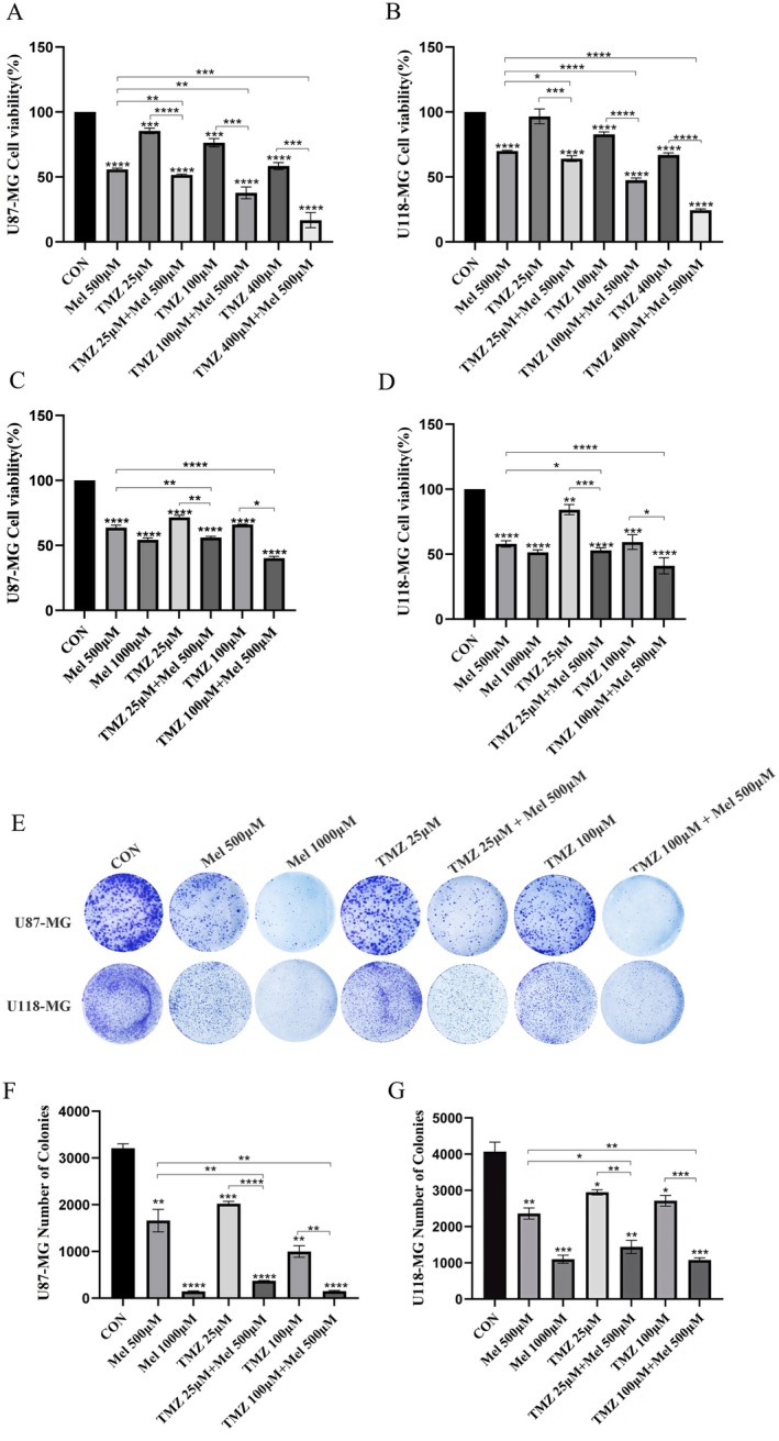
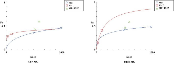
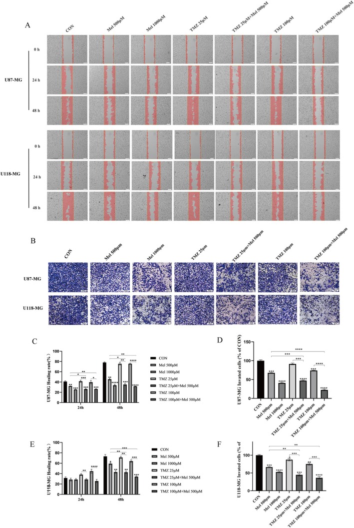
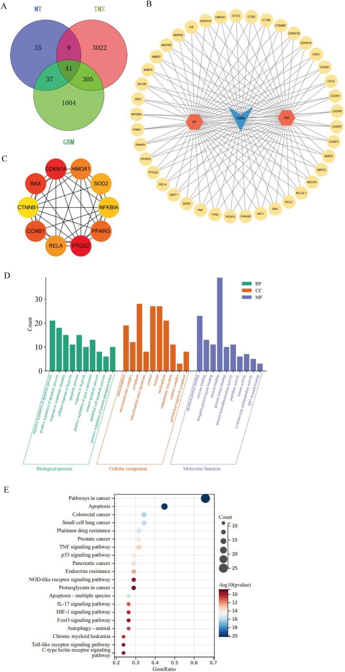
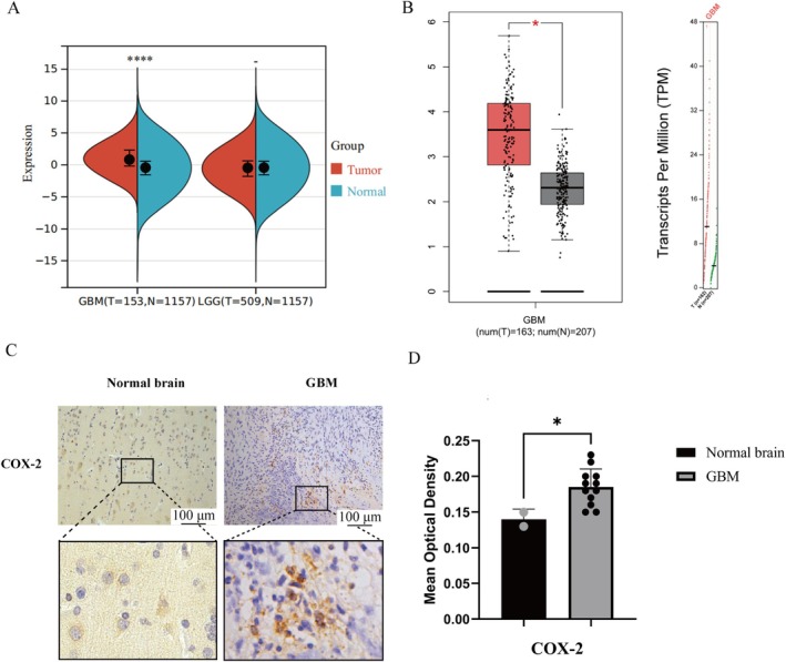
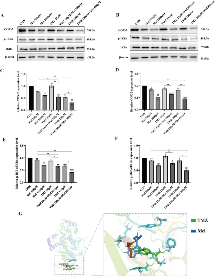
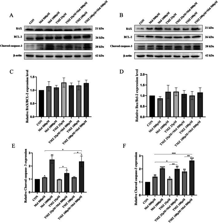
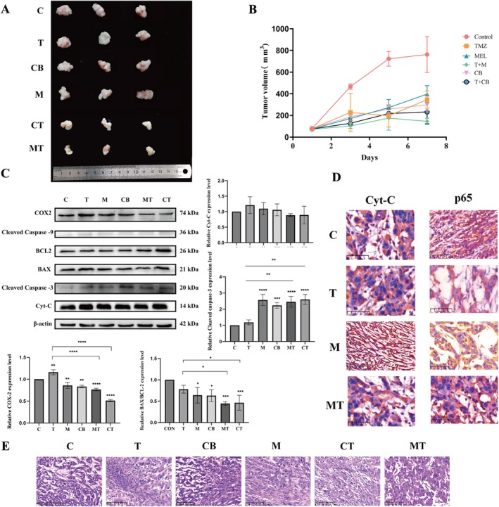
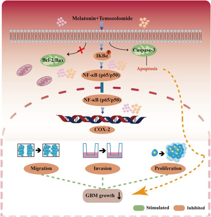
Glioblastoma (GBM) is an aggressive and highly malignant primary brain tumour, accounting for a significant proportion of adult brain tumours. It is associated with a poor prognosis and high recurrence rates. Although temozolomide (TMZ) remains the standard first-line chemotherapy for GBM, its clinical efficacy is often limited by the development of drug resistance and toxic effects on normal tissues. Melatonin (Mel), a natural indoleamine synthesised by the pineal gland, has demonstrated synergistic anti-tumour effects when combined with various chemotherapy agents in multiple studies. This study investigates the synergistic potential of Mel to enhance TMZ's therapeutic efficacy against GBM. The results demonstrate that the combination of Mel and TMZ significantly inhibits glioblastoma cell proliferation, migration, and invasion. Mechanistically, this synergistic effect is mediated through the NF-κB/COX-2 signalling pathway. Mel enhances TMZ's anti-tumour activity by inhibiting IκBα phosphorylation, suppressing NF-κB activation, and downregulating COX-2 expression. Additionally, the combination treatment induced apoptosis via activation of the Caspase-3 pathway. These results suggest that Mel can potentiate the therapeutic efficacy of TMZ in glioblastoma treatment, offering a promising strategy to overcome TMZ resistance while reducing its associated toxicity.

Keywords: NF‐κB/COX‐2; glioblastoma; melatonin; temozolomide.


Conflict of interest statement

The authors declare no conflicts of interest.

Figures

FIGURE 1
FIGURE 1
Effect of Mel and TMZ combination on cell viability and proliferation in GBM cells. (A, B) Human U87‐MG and U118‐MG cells were treated with Mel or TMZ or their combination at the indicated doses. At 48 h, the cell viability was measured using the MTT assay. Cell viability in the Mel 500 μM group was set as 100% reference. (C, D) U87‐MG and U118‐MG cells were treated with TMZ (25 μmol/L, 100 μmol/L) or Mel (500 μmol/L, 1000 μmol/L) alone or in combination. Colony formation of the GBM cells was captured in photographs (E), and the relative colony numbers were calculated (F, G). Data are presented as mean ± SD from three independent experiments. Statistical significance is indicated by *p < 0.05, **p < 0.01, ***p < 0.001, ****p < 0.0001.
FIGURE 2
FIGURE 2
Dose‐response curve for the individual drugs and combination of Mel and TMZ.
FIGURE 3
FIGURE 3
Effect of Mel and TMZ combination on cell migration and invasion in GBM cells. (A) Cell migration was analysed by a wound‐healing assay. The U87‐MG and U118‐MG cells were grown to full confluency, and the cell monolayers were wounded with a sterile pipette tip and washed with medium to remove detached cells from the plates (magnification 4×). Cells were then left either untreated or treated with Mel or TMZ alone or their combination. After 48 h, the wound gap was observed and photographed (magnification 10×). (C, E) The percentages of migrating cells were calculated relative to the original gap. (B) Cell invasion was analysed in GBM cells treated with indicated doses of TMZ or Mel alone or their combination for 48 h. Cell invasion was observed and photographed, and the percentages of invading cells (D, F) were calculated. The level of significance was indicated by *p < 0.05, **p < 0.01, ***p < 0.001 and ****p < 0.0001.
FIGURE 4
FIGURE 4
Network pharmacology was used to predict the targets and mechanism of Mel and TMZ in glioma. (A) Venn diagram of common targets of Mel, TMZ and GBM. (B) A network diagram of 41 Mel‐TMZ‐GBM interaction targets. (C) Top 10 hub genes based on the highest DMNC score, analysed by CytoHubba. (D) Gene Ontology (GO) analysis of common targets. BP, biological process; CC, cellular component; MF, molecular function. (E) Kyoto Encyclopedia of Genes and Genomes (KEGG) analysis of common targets.
FIGURE 5
FIGURE 5
The differential expression of COX‐2 in normal brain tissue and GBM through bioinformatics and immunohistochemistry. (A) The expression difference of COX‐2 in various gliomas and their normal brain tissues. (B) Differential COX‐2 expression within GBM and normal brain tissue. (C) Positive expression of COX‐2 (×200) expression in human GBM and normal brain tissue (magnification 10×). (D) Quantification of COX‐2 expression levels in GBM tissues. Staining intensity was assessed via average optical density (AOD) analysis. Results are presented as mean ± standard deviation (n = 12 GBM biopsies; n = 2 normal brain specimens). Statistical significance versus normal brain tissue *p < 0.05, and ****p < 0.0001.
FIGURE 6
FIGURE 6
Effect of Mel and TMZ combination on NF‐κB/COX‐2 signalling. U87‐MG cells and U118‐MG cells were treated with Mel (500 μM) or TMZ (25 μM, 100 μM) alone or their combination. (A, B) At 48 h after treatment, the protein levels of COX‐2, IKBα and p‐IKBα in U87‐MG and U118‐MG were detected by western blot analysis. (C) Statistical analysis of COX‐2, IKBα and p‐IKBα protein expression levels in U87‐MG cells by Mel synergistic TMZ. (D) Statistical analysis of COX‐2 protein expression levels in U118‐MG cells by Mel synergistic TMZ. (E) Statistical analysis of IKBα/p‐IKBα protein expression levels in U87‐MG cells by Mel synergistic TMZ. (F) Statistical analysis of IKBα/p‐IKBα protein expression levels in U118‐MG cells by Mel synergistic TMZ. (G) The three‐dimensional molecular docking structure of Mel and TMZ bound to COX‐2. n = 3 for each group. Statistical significance was defined as *p < 0.05, **p < 0.01, ***p < 0.001 and ****p < 0.0001.
FIGURE 7
FIGURE 7
Effect of Mel and TMZ combination on apoptosis‐related proteins. U87‐MG cells and U118‐MG cells were treated with Mel (500 μM) or TMZ (25 μM, 100 μM) alone or in combination. (A) At 48 h after treatment, the protein levels of BAX, Bcl‐2 and Cleaved caspase‐3 in U87‐MG were detected by western blot analysis. (B) At 48 h after treatment, the protein levels of BAX, Bcl‐2, and Cleaved caspase‐3 in U118‐MG were detected by western blot analysis. (C) Statistical analysis of BAX/Bcl‐2 protein expression levels in U87‐MG cells by Mel synergistic TMZ. (D) Statistical analysis of BAX/Bcl‐2 protein expression levels in U118‐MG cells by Mel synergistic TMZ. (E) Statistical analysis of Cleaved caspase‐3 protein expression levels in U87‐MG cells by Mel synergistic TMZ. (F) Statistical analysis of Cleaved caspase‐3 protein expression levels in U118‐MG cells by Mel synergistic TMZ. n = 3 for each group. Statistical significance was defined as *p < 0.05, **p < 0.01, ***p < 0.001 and ****p < 0.0001.
FIGURE 8
FIGURE 8
Research results on the therapeutic efficacy of Mel combined with TMZ in the GL261 subcutaneous xenograft mouse model. (A) Representative images of tumour. (B) Changes in tumour volume. (C) Expression profiles of apoptosis‐related proteins and COX2 protein in subcutaneous xenograft tumours. (D) Immunohistochemical localization images of p65 and Cyt‐C expression in xenograft tumours (magnification 4×). (E) HE staining of xenograft tumours. All data are presented as the mean ± SD derived from seven individual fields across six independent experimental replicates per treatment group (magnification 40×). (*p < 0.05, **p < 0.01, ***p < 0.001, ****p < 0.0001 vs. control group).
FIGURE 9
FIGURE 9
The mechanism of the action of Mel combined with TMZ in GBM cells.

Similar articles

References

    1. Schaff L. R. and Mellinghoff I. K., “Glioblastoma and Other Primary Brain Malignancies in Adults: A Review,” JAMA 329, no. 7 (2023): 574–587. - PMC - PubMed
    1. Vitovcova B., Skarkova V., Rudolf K., and Rudolf E., “Biology of Glioblastoma Multiforme‐Exploration of Mitotic Catastrophe as a Potential Treatment Modality,” International Journal of Molecular Sciences 21, no. 15 (2020): 5324. - PMC - PubMed
    1. Tan A. C., Ashley D. M., López G. Y., Malinzak M., Friedman H. S., and Khasraw M., “Management of Glioblastoma: State of the Art and Future Directions,” CA: A Cancer Journal for Clinicians 70, no. 4 (2020): 299–312. - PubMed
    1. Stepanovic A. and Nikitovic M., “Severe Hematologic Temozolomide‐Related Toxicity and Lifethreatening Infections,” Journal of B.U.ON.: Official Journal of the Balkan Union of Oncology 23, no. 1 (2018): 7–13. - PubMed
    1. Ortiz R., Perazzoli G., Cabeza L., et al., “Temozolomide: An Updated Overview of Resistance Mechanisms, Nanotechnology Advances and Clinical Applications,” Current Neuropharmacology 19, no. 4 (2021): 513–537. - PMC - PubMed

MeSH terms