

Magnetic Resonance Imaging monitoring of histotripsy effects in agar phantom
- PMID: 40804776
- PMCID: PMC12350826
- DOI: 10.1002/mp.18054
Magnetic Resonance Imaging monitoring of histotripsy effects in agar phantom
Abstract
Background: While the technical aspects of histotripsy have seen significant advancements, research on Magnetic Resonance Imaging (MRI) monitoring remains limited.
Purpose: This preliminary study explored the use of conventional T2-Weighted (T2-W) Turbo Spin Echo (TSE) imaging to monitor histotripsy lesions in a pure agar gel, as a potential research tool for MRI-guided histotripsy (MRgHt).
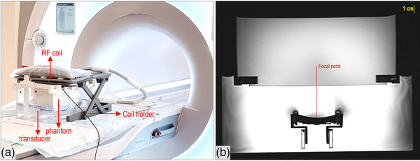
Methods: Histotripsy experiments were conducted in a 3T MRI scanner using a 2% weight per volume agar solution in water. Pulsed Focused Ultrasound (FUS) was applied at a duty factor of 2%, with a pulse repetition period of 1s, delivering up to 2000 pulses. T2-W TSE imaging was employed for intra-procedural monitoring with 10s updates and post-sonication assessment, with the first prioritizing rapid acquisition and the second focusing on resolution. Supplementary, MR thermometry tracked temperature variations throughout the procedure.
Results: Dynamic T2-W TSE imaging provided insights into the progress of phantom disruption. Mild thermal effects served as early signs of phantom response, as mechanical effects required more time to manifest. Typical histotripsy lesion characteristics were observed, including a hyperintense core with an elongated shape and a thin hypointense border. The developed temperatures remained well below hyperthermia levels, reaching a maximum of 34°C.
Conclusions: The presented methodology, utilizing a pure agar gel within a simple, versatile setup alongside conventional T2-W TSE imaging, holds promise for advancing MRgHt research.
Keywords: MRI; T2‐W; agar gel; histotripsy; lesion monitoring.
© 2025 The Author(s). Medical Physics published by Wiley Periodicals LLC on behalf of American Association of Physicists in Medicine.
Conflict of interest statement
The authors declare no conflicts of interest.
Figures


Similar articles
-
Phantom-based assessment of focused ultrasound thermal effects with conventional magnetic resonance imaging.Phys Med. 2025 Aug 23;137:105078. doi: 10.1016/j.ejmp.2025.105078. Online ahead of print. Phys Med. 2025. PMID: 40850157
-
Calibration correction to improve registration during cone-beam CT guided histotripsy.Med Phys. 2025 May;52(5):3216-3227. doi: 10.1002/mp.17644. Epub 2025 Jan 26. Med Phys. 2025. PMID: 39865624 Free PMC article.
-
MarkVCID cerebral small vessel consortium: II. Neuroimaging protocols.Alzheimers Dement. 2021 Apr;17(4):716-725. doi: 10.1002/alz.12216. Epub 2021 Jan 21. Alzheimers Dement. 2021. PMID: 33480157 Free PMC article.
-
The diagnostic accuracy and cost-effectiveness of magnetic resonance spectroscopy and enhanced magnetic resonance imaging techniques in aiding the localisation of prostate abnormalities for biopsy: a systematic review and economic evaluation.Health Technol Assess. 2013 May;17(20):vii-xix, 1-281. doi: 10.3310/hta17200. Health Technol Assess. 2013. PMID: 23697373 Free PMC article.
-
Contrast-enhanced ultrasound using SonoVue® (sulphur hexafluoride microbubbles) compared with contrast-enhanced computed tomography and contrast-enhanced magnetic resonance imaging for the characterisation of focal liver lesions and detection of liver metastases: a systematic review and cost-effectiveness analysis.Health Technol Assess. 2013 Apr;17(16):1-243. doi: 10.3310/hta17160. Health Technol Assess. 2013. PMID: 23611316 Free PMC article.
References
-
- Kisting MA, Jentink MS, Wagner MG, et al. Imaging for targeting, monitoring, and assessment after histotripsy: a non‐invasive, non‐thermal therapy for cancer. EMJ Radiology. doi: 10.33590/emjradiol/10308529 - DOI
MeSH terms
Substances
Grants and funding
LinkOut - more resources
Full Text Sources
Medical
