

Triamcinolone Acetonide-Assisted Visualization and Removal of Vitreous Cortex Remnants in Retinal Detachment: A Prospective Cohort Study
- PMID: 40804819
- PMCID: PMC12346414
- DOI: 10.3390/diagnostics15151854
Triamcinolone Acetonide-Assisted Visualization and Removal of Vitreous Cortex Remnants in Retinal Detachment: A Prospective Cohort Study
Abstract
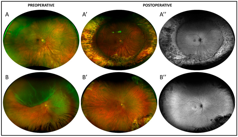
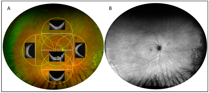
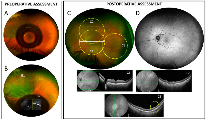
Background/Objectives: In rhegmatogenous retinal detachment (RRD), vitreous cortex remnants (VCRs) may contribute to the development and progression of proliferative vitreoretinopathy (PVR). This study aimed to evaluate potential toxicity and trauma secondary to VCRs visualization and removal during pars plana vitrectomy (PPV) for RRD. Methods: Prospective study on patients with primary RRD who underwent PPV. Imaging assessment included widefield OCT (WF-OCT), ultra-WF retinography and fundus autofluorescence (FAF). During PPV, a filtered and diluted triamcinolone acetonide (TA) solution (20 mg/mL) was used to evaluate the presence and extension of VCRs, removed using an extendible diamond-dusted sweeper (EDDS). After six months, retinal and retinal pigment epithelium toxicity and retinal trauma due to VCRs removal were investigated. Results: Retinal reattachment was achieved in 21/21 cases included in the study. No signs of retinal or RPE toxicity were detected and WF-OCT performed in the areas of VCRs removal revealed an intact inner retinal architecture in the majority of eyes, with minor and localized inner retinal indentations in 4 cases. Conclusions: VCRs visualization and removal using TA and EDDS appears to be safe, with no retinal toxicity and very limited and circumscribed mechanical trauma. This approach may contribute to reducing the risk of postoperative PVR.
Keywords: proliferative vitreoretinopathy; retinal toxicity; rhegmatogenous retinal detachment; triamcinolone acetonide; vitreous cortex remnants.
Conflict of interest statement
The authors declare no conflicts of interest.
Figures
Similar articles
-
Anti-vascular endothelial growth factor for prevention of postoperative vitreous cavity haemorrhage after vitrectomy for proliferative diabetic retinopathy.Cochrane Database Syst Rev. 2015 Aug 7;2015(8):CD008214. doi: 10.1002/14651858.CD008214.pub3. Cochrane Database Syst Rev. 2015. Update in: Cochrane Database Syst Rev. 2023 May 31;5:CD008214. doi: 10.1002/14651858.CD008214.pub4. PMID: 26250103 Free PMC article. Updated.
-
Pneumatic retinopexy versus scleral buckle for repairing simple rhegmatogenous retinal detachments.Cochrane Database Syst Rev. 2021 Nov 11;11(11):CD008350. doi: 10.1002/14651858.CD008350.pub3. Cochrane Database Syst Rev. 2021. PMID: 34762741 Free PMC article.
-
Pneumatic retinopexy versus scleral buckle for repairing simple rhegmatogenous retinal detachments.Cochrane Database Syst Rev. 2015 May 7;5(5):CD008350. doi: 10.1002/14651858.CD008350.pub2. Cochrane Database Syst Rev. 2015. Update in: Cochrane Database Syst Rev. 2021 Nov 11;11:CD008350. doi: 10.1002/14651858.CD008350.pub3. PMID: 25950286 Free PMC article. Updated.
-
Intravitreal methotrexate as an adjuvant in vitrectomy in cases of retinal detachment with proliferative vitreoretinopathy.Graefes Arch Clin Exp Ophthalmol. 2025 Feb;263(2):387-391. doi: 10.1007/s00417-024-06665-w. Epub 2024 Oct 17. Graefes Arch Clin Exp Ophthalmol. 2025. PMID: 39419842 Clinical Trial.
-
Pars Plana Vitrectomy with and without Supplemental Scleral Buckle for the Repair of Rhegmatogenous Retinal Detachment: A Meta-analysis.Ophthalmol Retina. 2022 Oct;6(10):871-885. doi: 10.1016/j.oret.2022.02.009. Epub 2022 Feb 26. Ophthalmol Retina. 2022. PMID: 35227949
References
-
- van Overdam K.A., van den Bosch T.P.P., van Etten P.G., Uppal G.S., Veckeneer M., Verdijk R.M. Novel insights into the pathophysiology of proliferative vitreoretinopathy: The role of vitreoschisis-induced vitreous cortex remnants. Acta Ophthalmol. 2022;100:e1749–e1759. doi: 10.1111/aos.15197. - DOI - PubMed
LinkOut - more resources
Full Text Sources
Research Materials
Miscellaneous
