

Deep Learning-Based Differentiation of Vertebral Body Lesions on Magnetic Resonance Imaging
- PMID: 40804827
- PMCID: PMC12345889
- DOI: 10.3390/diagnostics15151862
Deep Learning-Based Differentiation of Vertebral Body Lesions on Magnetic Resonance Imaging
Abstract
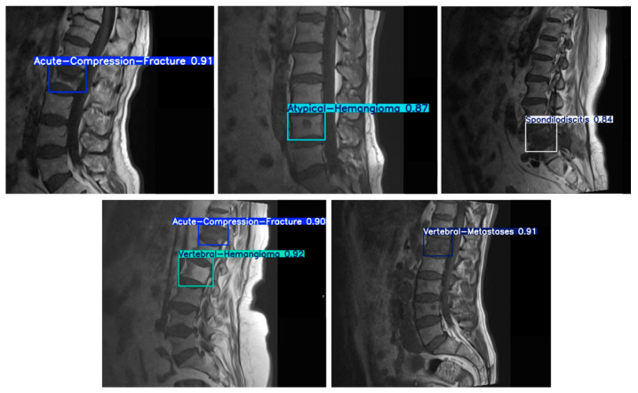
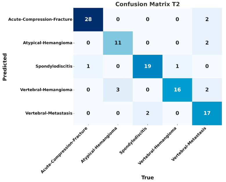
Objectives: Spinal diseases are commonly encountered health problems with a wide spectrum. In addition to degenerative changes, other common spinal pathologies include metastases and compression fractures. Benign tumors like hemangiomas and infections such as spondylodiscitis are also frequently observed. Although magnetic resonance imaging (MRI) is considered the gold standard in diagnostic imaging, the morphological similarities of lesions can pose significant challenges in differential diagnoses. In recent years, the use of artificial intelligence applications in medical imaging has become increasingly widespread. In this study, we aim to detect and classify vertebral body lesions using the YOLO-v8 (You Only Look Once, version 8) deep learning architecture. Materials and Methods: This study included MRI data from 235 patients with vertebral body lesions. The dataset comprised sagittal T1- and T2-weighted sequences. The diagnostic categories consisted of acute compression fractures, metastases, hemangiomas, atypical hemangiomas, and spondylodiscitis. For automated detection and classification of vertebral lesions, the YOLOv8 deep learning model was employed. Following image standardization and data augmentation, a total of 4179 images were generated. The dataset was randomly split into training (80%) and validation (20%) subsets. Additionally, an independent test set was constructed using MRI images from 54 patients who were not included in the training or validation phases to evaluate the model's performance. Results: In the test, the YOLOv8 model achieved classification accuracies of 0.84 and 0.85 for T1- and T2-weighted MRI sequences, respectively. Among the diagnostic categories, spondylodiscitis had the highest accuracy in the T1 dataset (0.94), while acute compression fractures were most accurately detected in the T2 dataset (0.93). Hemangiomas exhibited the lowest classification accuracy in both modalities (0.73). The F1 scores were calculated as 0.83 for T1-weighted and 0.82 for T2-weighted sequences at optimal confidence thresholds. The model's mean average precision (mAP) 0.5 values were 0.82 for T1 and 0.86 for T2 datasets, indicating high precision in lesion detection. Conclusions: The YOLO-v8 deep learning model we used demonstrates effective performance in distinguishing vertebral body metastases from different groups of benign pathologies.
Keywords: MRI; YOLO-v8; classification; deep learning; detection; metastasis; vertebral body lesions.
Conflict of interest statement
The authors declare no conflicts of interest related to this study.
Figures


Similar articles
-
Prescription of Controlled Substances: Benefits and Risks.2025 Jul 6. In: StatPearls [Internet]. Treasure Island (FL): StatPearls Publishing; 2025 Jan–. 2025 Jul 6. In: StatPearls [Internet]. Treasure Island (FL): StatPearls Publishing; 2025 Jan–. PMID: 30726003 Free Books & Documents.
-
A deep learning approach to direct immunofluorescence pattern recognition in autoimmune bullous diseases.Br J Dermatol. 2024 Jul 16;191(2):261-266. doi: 10.1093/bjd/ljae142. Br J Dermatol. 2024. PMID: 38581445
-
Comparison of Two Modern Survival Prediction Tools, SORG-MLA and METSSS, in Patients With Symptomatic Long-bone Metastases Who Underwent Local Treatment With Surgery Followed by Radiotherapy and With Radiotherapy Alone.Clin Orthop Relat Res. 2024 Dec 1;482(12):2193-2208. doi: 10.1097/CORR.0000000000003185. Epub 2024 Jul 23. Clin Orthop Relat Res. 2024. PMID: 39051924
-
A systematic review of evidence on malignant spinal metastases: natural history and technologies for identifying patients at high risk of vertebral fracture and spinal cord compression.Health Technol Assess. 2013 Sep;17(42):1-274. doi: 10.3310/hta17420. Health Technol Assess. 2013. PMID: 24070110 Free PMC article.
-
Contrast-enhanced ultrasound using SonoVue® (sulphur hexafluoride microbubbles) compared with contrast-enhanced computed tomography and contrast-enhanced magnetic resonance imaging for the characterisation of focal liver lesions and detection of liver metastases: a systematic review and cost-effectiveness analysis.Health Technol Assess. 2013 Apr;17(16):1-243. doi: 10.3310/hta17160. Health Technol Assess. 2013. PMID: 23611316 Free PMC article.
References
Grants and funding
LinkOut - more resources
Full Text Sources
