Deep Learning-Based Recurrence Prediction in HER2-Low Breast Cancer: Comparison of MRI-Alone, Clinicopathologic-Alone, and Combined Models
- PMID: 40804859
- PMCID: PMC12346550
- DOI: 10.3390/diagnostics15151895
Deep Learning-Based Recurrence Prediction in HER2-Low Breast Cancer: Comparison of MRI-Alone, Clinicopathologic-Alone, and Combined Models
Abstract
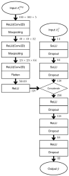
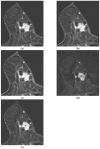
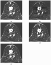
Background/Objectives: To develop a DL-based model predicting recurrence risk in HER2-low breast cancer patients and to compare performance of the MRI-alone, clinicopathologic-alone, and combined models. Methods: We analyzed 453 patients with HER2-low breast cancer who underwent surgery and preoperative breast MRI between May 2018 and April 2022. Patients were randomly assigned to either a training cohort (n = 331) or a test cohort (n = 122). Imaging features were extracted from DCE-MRI and ADC maps, with regions of interest manually annotated by radiologists. Clinicopathological features included tumor size, nodal status, histological grade, and hormone receptor status. Three DL prediction models were developed: a CNN-based MRI-alone model, a clinicopathologic-alone model based on a multi-layer perceptron (MLP) and a combined model integrating CNN-extracted MRI features with clinicopathological data via MLP. Model performance was evaluated using AUC, sensitivity, specificity, and F1-score. Results: The MRI-alone model achieved an AUC of 0.69 (95% CI, 0.68-0.69), with a sensitivity of 37.6% (95% CI, 35.7-39.4), specificity of 87.5% (95% CI, 86.9-88.2), and F1-score of 0.34 (95% CI, 0.33-0.35). The clinicopathologic-alone model yielded the highest AUC of 0.92 (95% CI, 0.92-0.92) and sensitivity of 93.6% (95% CI, 93.4-93.8), but showed the lowest specificity (72.3%, 95% CI, 71.8-72.8) and F1-score of 0.50 (95% CI, 0.49-0.50). The combined model demonstrated the most balanced performance, achieving an AUC of 0.90 (95% CI, 0.89-0.91), sensitivity of 80.0% (95% CI, 78.7-81.3), specificity of 83.2% (95% CI: 82.7-83.6), and the highest F1-score of 0.55 (95% CI, 0.54-0.57). Conclusions: The DL-based model combining MRI and clinicopathological features showed superior performance in predicting recurrence in HER2-low breast cancer. This multimodal approach offers a framework for individualized risk assessment and may aid in refining follow-up strategies.
Keywords: breast neoplasm; deep learning; human epidermal growth factor receptor 2; prediction algorithms; recurrence.
Conflict of interest statement
The authors declare no conflicts of interest.
Figures


Similar articles
-
Development of a prediction model for HER2 low breast cancer using quantitative intra- and peri-tumoral heterogeneity and MRI features on high-spatial resolution ultrafast DCE-MRI.Quant Imaging Med Surg. 2025 Sep 1;15(9):7788-7802. doi: 10.21037/qims-24-976. Epub 2025 Aug 18. Quant Imaging Med Surg. 2025. PMID: 40893528 Free PMC article.
-
Comparison of Two Modern Survival Prediction Tools, SORG-MLA and METSSS, in Patients With Symptomatic Long-bone Metastases Who Underwent Local Treatment With Surgery Followed by Radiotherapy and With Radiotherapy Alone.Clin Orthop Relat Res. 2024 Dec 1;482(12):2193-2208. doi: 10.1097/CORR.0000000000003185. Epub 2024 Jul 23. Clin Orthop Relat Res. 2024. PMID: 39051924
-
Development and validation of an MRI spatiotemporal interaction model for early noninvasive prediction of neoadjuvant chemotherapy response in breast cancer: a multicentre study.EClinicalMedicine. 2025 Jun 12;85:103298. doi: 10.1016/j.eclinm.2025.103298. eCollection 2025 Jul. EClinicalMedicine. 2025. PMID: 40584836 Free PMC article.
-
Artificial intelligence for diagnosing exudative age-related macular degeneration.Cochrane Database Syst Rev. 2024 Oct 17;10(10):CD015522. doi: 10.1002/14651858.CD015522.pub2. Cochrane Database Syst Rev. 2024. PMID: 39417312
-
Magnetic resonance perfusion for differentiating low-grade from high-grade gliomas at first presentation.Cochrane Database Syst Rev. 2018 Jan 22;1(1):CD011551. doi: 10.1002/14651858.CD011551.pub2. Cochrane Database Syst Rev. 2018. PMID: 29357120 Free PMC article.
References
-
- Ramakrishna N., Temin S., Chandarlapaty S., Crews J.R., Davidson N.E., Esteva F.J., Giordano S.H., Kirshner J.J., Krop I.E., Levinson J., et al. Recommendations on Disease Management for Patients With Advanced Human Epidermal Growth Factor Receptor 2-Positive Breast Cancer and Brain Metastases: ASCO Clinical Practice Guideline Update. J. Clin. Oncol. 2018;36:2804–2807. doi: 10.1200/JCO.2018.79.2713. - DOI - PubMed
-
- Giordano S.H., Franzoi M.A.B., Temin S., Anders C.K., Chandarlapaty S., Crews J.R., Kirshner J.J., Krop I.E., Lin N.U., Morikawa A., et al. Systemic Therapy for Advanced Human Epidermal Growth Factor Receptor 2-Positive Breast Cancer: ASCO Guideline Update. J. Clin. Oncol. 2022;40:2612–2635. doi: 10.1200/JCO.22.00519. - DOI - PubMed
-
- Wolff A.C., Somerfield M.R., Dowsett M., Hammond M.E.H., Hayes D.F., McShane L.M., Saphner T.J., Spears P.A., Allison K.H. Human Epidermal Growth Factor Receptor 2 Testing in Breast Cancer: ASCO-College of American Pathologists Guideline Update. J. Clin. Oncol. 2023;41:3867–3872. doi: 10.1200/JCO.22.02864. - DOI - PubMed
LinkOut - more resources
Full Text Sources
Research Materials
Miscellaneous

