Multimodal Imaging Detection of Difficult Mammary Paget Disease: Dermoscopy, Reflectance Confocal Microscopy, and Line-Field Confocal-Optical Coherence Tomography
- PMID: 40804863
- PMCID: PMC12346694
- DOI: 10.3390/diagnostics15151898
Multimodal Imaging Detection of Difficult Mammary Paget Disease: Dermoscopy, Reflectance Confocal Microscopy, and Line-Field Confocal-Optical Coherence Tomography
Abstract
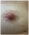
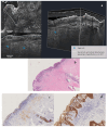
Mammary Paget disease (MPD) is a rare cutaneous malignancy associated with underlying ductal carcinoma in situ (DCIS) or invasive ductal carcinoma (IDC). Clinically, it appears as eczematous changes in the nipple and areola complex (NAC), which may include itching, redness, crusting, and ulceration; these symptoms can sometimes mimic benign dermatologic conditions such as nipple eczema, making early diagnosis challenging. A 56-year-old woman presented with persistent erythema and scaling of the left nipple, which did not respond to conventional dermatologic treatments: a high degree of suspicion prompted further investigation. Reflectance confocal microscopy (RCM) revealed atypical, enlarged epidermal cells with irregular boundaries, while line-field confocal-optical coherence tomography (LC-OCT) demonstrated thickening of the epidermis, hypo-reflective vacuous spaces and abnormally large round cells (Paget cells). These non-invasive imaging findings were consistent with an aggressive case of Paget disease despite the absence of clear mammographic evidence of underlying carcinoma: in fact, several biopsies were needed, and at the end, massive surgery was necessary. Non-invasive imaging techniques, such as dermoscopy, RCM, and LC-OCT, offer a valuable diagnostic tool in detecting Paget disease, especially in early stages and atypical forms.
Keywords: dermoscopy; line-field optical-coherence tomography; mammary Paget disease; nipple areola complex; reflectance confocal microscopy.
Conflict of interest statement
The authors declare no conflicts of interest.
Figures


References
-
- Scott-Emuakpor R., Reza-Soltani S., Altaf S., NR K., Kołodziej F., Sil-Zavaleta S., Nalla M., Ullah M.N., Qureshi M.R., Ahmadi Y., et al. Mammary Paget’s Disease Mimicking Benign and Malignant Dermatological Conditions: Clinical Challenges and Diagnostic Considerations. Cureus. 2024;16:e65378. doi: 10.7759/cureus.65378. - DOI - PMC - PubMed
-
- Apalla Z., Errichetti E., Kyrgidis A., Stolz W., Puig S., Malvehy J., Zalaudek I., Moscarella E., Longo C., Blum A., et al. Dermoscopic features of mammary Paget’s disease: A retrospective case-control study by the International Dermoscopy Society. J. Eur. Acad. Dermatol. Venereol. 2019;33:1892–1898. doi: 10.1111/jdv.15732. - DOI - PubMed
LinkOut - more resources
Full Text Sources

