

Texture and Color Enhancement Imaging-Assisted Endocytoscopy Improves Characterization of Gastric Precancerous Conditions: A Set of Interesting Comparative Images
- PMID: 40804890
- PMCID: PMC12346551
- DOI: 10.3390/diagnostics15151925
Texture and Color Enhancement Imaging-Assisted Endocytoscopy Improves Characterization of Gastric Precancerous Conditions: A Set of Interesting Comparative Images
Abstract
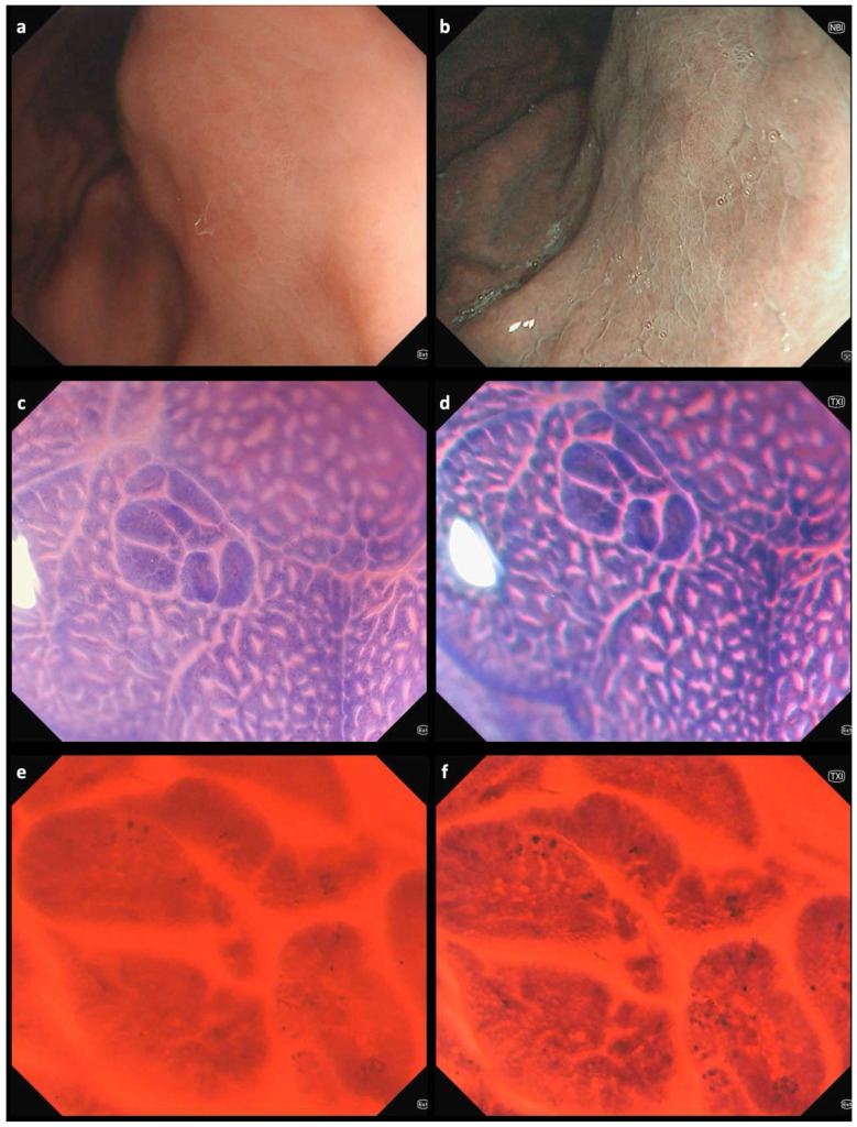
Chronic atrophic gastritis and intestinal metaplasia (IM) are gastric precancerous conditions (GPCs) associated with an increased risk of gastric cancer. Early detection and accurate characterization of GPC are therefore crucial for risk stratification and the implementation of preventive strategies. In the absence of clear mucosal changes observed through white-light imaging (WLI) or virtual chromoendoscopy, endocytoscopy can help unveil the presence of GPC by enabling in vivo assessment of nuclear and cellular structures at ultra-high magnification. Endocytoscopy is typically performed using WLI following dye-based staining of the mucosa. In this case, we demonstrate that combining endocytoscopy with the texture and color enhancement imaging (TXI) mode substantially improves the assessment of the gastric mucosa. In a 61-year-old man undergoing esophagogastroduodenoscopy, WLI showed multifocal erythema in the stomach, without clearly visible lesions on either WLI or narrow-band imaging. Conventional endocytoscopy revealed multiple small spots of IM with characteristic changes in glandular structures, which were even more evident when using the TXI mode. Histological analysis of targeted biopsies confirmed small foci of IM in both the antrum and corpus. The patient was enrolled in a surveillance program because of his clinical background. The combination of endocytoscopy with the TXI mode significantly enhances the delineation of mucosal and cellular architecture, supporting a more accurate optical diagnosis.
Keywords: endocytoscopy; gastric atrophy; gastric cancer; intestinal metaplasia; preneoplastic lesions; stomach.
Conflict of interest statement
The authors declare no conflicts of interest.
Figures
Similar articles
-
Prescription of Controlled Substances: Benefits and Risks.2025 Jul 6. In: StatPearls [Internet]. Treasure Island (FL): StatPearls Publishing; 2025 Jan–. 2025 Jul 6. In: StatPearls [Internet]. Treasure Island (FL): StatPearls Publishing; 2025 Jan–. PMID: 30726003 Free Books & Documents.
-
Comparison of Effective Imaging Modalities for Detecting Gastric Neoplasms: A Randomized 3-Arm Phase II Trial.Am J Gastroenterol. 2024 Oct 1;119(10):2010-2018. doi: 10.14309/ajg.0000000000002871. Epub 2024 May 16. Am J Gastroenterol. 2024. PMID: 38752623 Free PMC article. Clinical Trial.
-
Signs and symptoms to determine if a patient presenting in primary care or hospital outpatient settings has COVID-19.Cochrane Database Syst Rev. 2022 May 20;5(5):CD013665. doi: 10.1002/14651858.CD013665.pub3. Cochrane Database Syst Rev. 2022. PMID: 35593186 Free PMC article.
-
Endocytoscopy in real-time assessment of histological and endoscopic activity in ulcerative colitis.World J Gastrointest Endosc. 2025 Jul 16;17(7):108082. doi: 10.4253/wjge.v17.i7.108082. World J Gastrointest Endosc. 2025. PMID: 40677581 Free PMC article.
-
MRI software and cognitive fusion biopsies in people with suspected prostate cancer: a systematic review, network meta-analysis and cost-effectiveness analysis.Health Technol Assess. 2024 Oct;28(61):1-310. doi: 10.3310/PLFG4210. Health Technol Assess. 2024. PMID: 39367754 Free PMC article.
References
-
- Kurtcehajic A., Zerem E., Bokun T., Alibegovic E., Kunosic S., Hujdurovic A., Tursunovic A., Ljuca K. Could near focus endoscopy, narrow-band imaging, and acetic acid improve the visualization of microscopic features of stomach mucosa? World J. Gastrointest. Endosc. 2024;16:157–167. doi: 10.4253/wjge.v16.i3.157. - DOI - PMC - PubMed
Grants and funding
LinkOut - more resources
Full Text Sources
